Electromyography
Electromyography
A signal is, by definition, nothing more than the graphical representation of the temporal trend of a physical quantity. In the case of the surface electromyogram (sEMG), this quantity is the potential difference generated by the muscle during its contraction, which produces an electric current in the tissues and a potential difference that is ultimately recorded on the skin. The graphical representation of this is the electromyogram or electromyographic trace or electromyographic signal or sEMG.
When detecting and recording an sEMG signal, two main aspects that influence the fidelity of the recording must be considered: the signal-to-noise ratio and distortion. The first is defined as the ratio between the energy of the useful signal (i.e., the desired signal) and the energy of the noise. The latter consists not only of actual noise (which we could imagine as the background hiss of old 78 RPM records) but also of any other signal that is simply unwanted, such as cardiac signals, signals from other muscles, or signals due to artifacts. This contamination, although often referred to as noise, should more accurately be termed interference, leaving the term noise for purely thermal noise. Distortion, on the other hand, is an alteration of the useful sEMG waveform that manifests mathematically as an undesired variation in the frequency components of the sEMG signal.
The signal-to-noise ratio and distortion are two problems that, by altering the recorded signal representation, can modify or hide the information the sEMG signal is meant to convey.
Noise Characteristics of sEMG: Electromagnetic Interference: Noise sources include electromagnetic environmental noise, such as 50 Hz power line interference, often exceeding the sEMG signal amplitude by several magnitudes. Proper differential amplifier design and shielding are crucial to reducing this interference.
Movement Artifacts: Artifacts arise from electrode movement or cable displacement during recording. Flexible electrodes and shielded cables minimize these disturbances, which typically affect frequencies below 20 Hz and can be filtered out without impacting the useful signal band (20–500 Hz).
Intrinsic Randomness: The sEMG signal's quasi-random nature, attributed to motor unit discharge variability, adds complexity. While low-frequency components (0–20 Hz) often reflect noise, high-frequency analysis provides clinically meaningful insights.
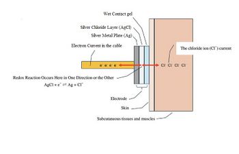
Electrodes: Electrodes serve as an interface, converting ionic currents in tissues into electronic signals. Commonly used Ag/AgCl electrodes are reversible and consumable, enabling consistent measurements while avoiding polarization. Proper placement and maintenance are essential for minimizing liquid junction potentials and ensuring signal fidelity.
Amplifier Design: Differential Amplification and CMRR: Differential amplifiers enhance the difference between two electrode inputs while suppressing common-mode noise, such as power line interference. The Common-Mode Rejection Ratio (CMRR) quantifies this efficiency, with optimal designs achieving values of 90–120 dB.
Input Impedance: High input impedance minimizes signal loss and distortion, ensuring accurate recording. Modern amplifiers achieve input impedances of up to 1015 ohms, reducing electrode load and preserving signal integrity.
Signal Processing: Traditional methods like rectification and integration are supplemented by modern techniques, including Root Mean Square (RMS) computation, which provides a robust measure of muscle activity power. Time-domain measurements, such as latency and conduction velocity, are critical for clinical and biomechanical analyses.
Conclusions: This chapter underscores the critical factors affecting sEMG fidelity, from electrode design to amplifier characteristics and signal processing methods. While challenges such as noise and distortion persist, advancements in technology and methodology enable precise muscle activity analysis, fostering enhanced diagnostic and research applications.
To read the full text of this chapter, log in or request an account
A Google Account is needeed to request a Member Account
