Artificial Neural Networks: Automatic Neuromuscular Diagnostic
Artificial Neural Networks: Automatic Neuromuscular Diagnostic
Article by Neri Accornero · Antonietta Romaniello · Giancarlo Filligoi · Edvina Galiè · Bruno Gregori
|
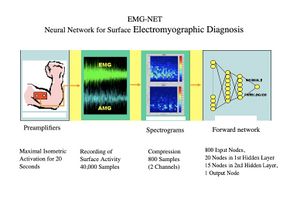
A preliminary study is presented on the automatic diagnosis of neuromuscular disorders using artificial neural networks that evaluate electrical and acoustic surface signals of the muscles under examination. The analysis of the electrical and acoustic (vibratory) activity captured on the muscle surface proves particularly promising for the non-invasiveness of the method, compared to the traditional electromyographic (EMG) exam performed with a disposable needle electrode, for its low cost, and because the recorded activity, coming from a much larger muscle mass than that explored with a needle electrode, theoretically allows for the detection of even subclinical pathologies. Surface recording of muscle activity is also particularly advantageous in sports medicine, in neuromotor rehabilitation, and in the functional control of motorized prostheses. Vibratory signals produced by the contraction and sliding of individual muscle fibers can also be derived from the muscle surface. This activity, called an acousto-myogram (AMG), provides relevant information that correlates with the force exerted and fatigue. Some recent studies have documented the complementarity of the two signals, the electrical and the acoustic, in the evaluation of exercise and muscle fatigue. Surface signals are inherently complex, in the mathematical sense, as they are composed of the sum of the activities of numerous sources (muscle fibers) that are not strictly correlated. Traditional analysis methods, using descriptors such as amplitude, spectrum, and morphology, are difficult to apply. Similarly, the use of expert diagnostic systems, based on rules, has proven to be ineffective. In this context, connectionist systems or artificial neural networks seem particularly suitable, as they achieve remarkable classification capabilities by being trained "by example" rather than by rules. To evaluate this method, we developed a system aimed at performing a screening between normal and pathological conditions in the context of neuromuscular disorders. (Fig. 1)
Considering the particularly encouraging results, a second version of the system was developed, aimed at differentiating within pathological cases between myopathies and neuropathies. This is currently under evaluation in a sample of patients with neuromuscular pathologies in the orofacial region.
Conclusions: After achieving a good level of training on the example case set (error = 0.03), a test was conducted on the 30 randomly selected test cases. The results were decidedly encouraging, as all new cases were correctly classified. Subsequent tests were carried out using only the electromyographic data and only the acousto-myographic data, with the observation that the network predominantly uses the information contained in the EMG signal (>90%) and that the contribution of the acoustic signal, although consistently low (<10%), complements the diagnostic accuracy. (Fig. 4).
From this preliminary study, the following conclusions can be drawn:
- The information contained in surface electrical and acoustic signals is sufficient to diagnose normality or pathology.
- The surface electromyographic signal appears indispensable for classification, but the correlation with the acoustic signal improves diagnostic accuracy.
- A connectionist system can perform a correct diagnostic classification using a significant compression of the aforementioned signals, consisting of the spectrograms of the two 20-second signals, segmented into 1-second epochs with 20 frequency levels, thus compressing the information from 40,000 samples to 800.
- The network's analog output, with values ranging between +1 and -1, allows for a “quantitative” assessment of the diagnosis of normality or pathology.
These results encourage the continuation of the study with the aim of further articulating the diagnostic classification, discriminating between neuropathies and myopathies within the pathological group. To this end, it will be necessary to significantly expand the set of example cases with pathologies from the two mentioned categories, carefully diagnosed clinically and electromyographically.
To read the full text of this chapter, log in or request an account
A Google Account is needeed to request a Member Account
