Stimolazione Elettrica e Magnetica del Sistema Nervoso Centrale e Periferico: Modellizzazione dei Campi Generati e Interpretazione dei Dati
Stimolazione Elettrica e Magnetica del Sistema Nervoso Centrale e Periferico: Modellizzazione dei Campi Generati e Interpretazione dei Dati
Article by Paolo Ravazzani · Gabriella Tognola · Marta Parazzini · Vincenzo Raschellà · Ferdinando Grandori
|
Introduction
Magnetic stimulation of the central and peripheral nervous system can now be considered a common method in clinical neurophysiology for assessing the conduction status of motor efferent pathways and peripheral nerves. Introduced in the mid-1980s as an improvement over direct electrical stimulation, it is based on the application of rapidly changing and high-intensity magnetic fields (up to 2 T), which induce an electric field in brain and nerve tissues through electromagnetic induction [1].
In a short time, this technique has spread widely, becoming an excellent clinical tool for evaluating the functionality of motor efferent pathways and diagnosing central nervous system dysfunctions. Despite this, the level of empiricism in the entire method remains significant, and many areas of investigation remain open, both in the technological, neurophysiological, and clinical fields.
The first scientific studies documenting the behavior of the induced electric field by different types of stimulators appeared in the literature around 10-15 years ago, with the development of numerous mathematical models, both analytical and numerical [2].
Despite these studies, technological progress related to the improvement and innovation of stimulators and stimulation coils has been scarce and often limited to changes in coil shape. In recent years, stimulation devices have not seen significant progress. In particular, there have been few improvements in focusing and controlling the induced electric field, while only systems for rapid repetitive stimulation have seen some innovations.
The ability to control the focusing of the induced electric field by a coil system would greatly expand the application areas of magnetic stimulation. For example, one could consider the potential offered by stimulating nerve centers responsible for controlling respiratory muscles (for patients with descending tract lesions), studying new methods of ventricular defibrillation, and developing non-invasive temporary pacemaking techniques.
For these interesting research prospects, it is essential, in addition to optimizing the construction of the coils and associated equipment, to search for new configurations with greater field focusing capabilities. In fact, the ability to focus the induced electric field in arbitrarily small regions, and consequently to concentrate induced currents in these regions, remains a decisive aspect for a qualitative leap in the use of magnetic stimulation.
This contribution aims to provide a brief overview of the current state of magnetic stimulation from a methodological and technological perspective and to introduce and discuss some innovative approaches that could contribute to the future development of the method and its use in new application fields.
Non-invasive Stimulation of the Motor Cortex
Transcranial Electrical Stimulation
In the past 20 years, the first attempts to stimulate the motor cortex were achieved through electrical stimulation. In 1980, Merton and Morton [3] developed a stimulation method using two electrodes placed on the scalp, near the motor area. This method is called bipolar because only two electrodes are used: an anode and a cathode. A step stimulus pulse is sent to the anode, with a very rapid rise time and voltage levels between 800 and 1800 V, with a current at the electrode of around 100 mA and a duration between 10 and 50 μs.
Due to the non-homogeneous nature of the head structures and brain tissues, electrical conductivity varies greatly from tissue to tissue. Some of the current is sufficient to stimulate the target motor structures, but most of it affects the scalp, causing the subject an almost unbearable painful sensation. This was considered the main disadvantage of this method and the primary reason for its limited use and eventual abandonment by the mid-1980s.
Transcranial Magnetic Stimulation
Magnetic stimulation is a method that overcomes the main drawbacks of electrical stimulation and is now considered one of the most widely used tools in clinical neurophysiology, particularly for diagnosing central and peripheral nervous system disorders. The main characteristic of this method is its ability to stimulate nerve structures such as the motor cortex or spinal nerve roots non-invasively and without causing any painful sensations to the subject.
Cortical magnetic stimulation, in its current form, was first proposed by Barker and collaborators in 1985, [1] who achieved it using short monophasic magnetic pulses with a peak amplitude of up to 2 T, then recording the motor response produced.
Transcranial Magnetic Stimulation: The Stimulator
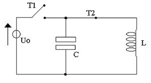
Simplified diagram of a stimulator circuit. The typical circuit parameters are chosen as L = 20 μH and C = 100 μF.
The typical structure of a stimulation device consists of two parts (Fig. 1):
a current pulse generator, which can produce peak currents exceeding 5 kA, and a stimulation coil, through which strong magnetic pulses are generated, with peak values reaching around 2 T and durations of about 300 μs. A trigger signal controls the discharge of energy stored in the capacitor into the stimulation coil. The pulse is controlled by a thyristor, which allows high currents to pass through in just a few microseconds, generating a monophasic discharge current with minimal reverse current. Additionally, through a small control current that manages its switching, currents in the range of several kA can flow.
The discharge current flows through the stimulation coil, generating a variable magnetic field that, in turn, induces activation currents within the tissues. Since the windings have a non-negligible resistance, energy dissipation due to Joule heating, with consequent coil heating, becomes significant.
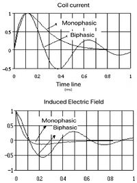
The induced currents within the human body, if of adequate amplitude and duration, can stimulate neuromuscular tissue similarly to electrical stimulation. The use of monophasic currents reduces heat dissipation in the coil, the noise associated with its rapid mechanical deformation (clicking sound), and artifacts, thereby increasing the accuracy of stimulation. Moreover, the monophasic pulse is more stable and defined, allowing for better interpretation of the responses (Fig. 2).
Repetitive Stimulation
Repetitive stimulation is based on the same principle as classical magnetic stimulation (single pulse). To achieve high stimulation frequencies (up to 50 Hz), smaller capacitors with shorter charge and discharge times are required compared to single-pulse magnetic stimulation. For this purpose, a series of capacitors activated in sequence by a microprocessor through a trigger signal are used, all discharging into the same stimulation coil. One of the main challenges in this case is managing the heating of the coils due to Joule heating. This imposes limits on the frequency of the stimuli and the duration during which the stimulus can be maintained at maximum intensity.
Focusing and Control of Induced Fields
Focusing the electric fields induced within biological tissues through magnetic stimulation must undoubtedly be considered one of the priority aspects in improving the technique. Here, focusing refers to the ability to concentrate an effective electric field—a field capable of stimulating excitable tissues such as nerve or muscle tissue—on a small region. In practice, focusing the induced electric field in cortical stimulation means selectively stimulating a set of neurons so that detectable effects on the motor system, sensory system, or cognitive activity can be observed.
However, the complexity of brain structure and the numerous synaptic contacts along the efferent and afferent pathways to the cerebral cortex do not allow for a linear relationship between the electromagnetic stimulus and the desired response. Additionally, the stimulation thresholds of different sets of neurons vary depending on the type of neuron, the depth from the cortical surface, and the orientation of nerve structures, making both field intensity and direction important. Moreover, attempting to concentrate the induced electric fields in a small brain region produces low-intensity fields. Thus, focusing effective fields seems to be a compromise between the area where the field exceeds a certain threshold and the maximum achievable field intensity.
A similar but distinct parameter from focusing is stimulation efficiency. This concept is more related to energy considerations for stimulation coils, in the sense of determining the minimum energy required for stimulation and how localized the resulting stimulus can be. It is conceivable that future developments in the technique will synthesize these two operational and design objectives, but as it stands, due to the fundamental physics underlying magnetic stimulation, it can be stated that focusing the electric field in a specific region of the body does not, at least theoretically, equate to minimizing the required energy—i.e., the energy that must be stored in the coil to generate the time-varying magnetic pulse.
Finally, an additional aspect related to focusing is the ability to concentrate the fields in depth and not just on a superficial region. To emphasize this aspect, we will refer to surface focusing and deep or volumetric focusing.
A stimulation coil is made up of one or more well-insulated copper windings, along with other electronic devices such as temperature sensors and safety switches [4]. Circular coils are the simplest and most straightforward design, and to this day, they are the most commonly used for stimulating the central nervous system (CNS) or spinal nerve roots. In clinical neurology, the CNS is stimulated with a circular coil of relatively large diameter, and the coil's position and angle relative to the subject’s head are adjusted to obtain the desired motor response.
The expression for the induced electric field inside a spherical medium has the following form:
In practice, the induced electric field is directly proportional to the time derivative of the current in the coil and to a spatial factor , which depends on the shapes and dimensions of the media and the coil, their electrical properties, and the relative position of the coil with respect to the point where the field is being calculated . The temporal behavior of is therefore proportional only to the time derivative of the current passing through the coil, and thus it depends on the type of current pulse (e.g., monophasic or biphasic) delivered by the stimulator to the coil (Fig. 2). As a result, the study of field focusing is closely related to the spatial factor .
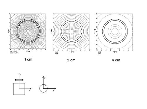
Now, let us consider a circular coil placed tangentially, in such a way that a radius of a sphere, ideally representing the head, coincides with the coil’s axis. The induced electric field (Fig. 3) is zero along the coil’s axis of symmetry and reaches its maximum just below the coil’s edge [5].
In practice, it is possible to obtain a maximum induced electric field only in regions close to the coil’s edge. Furthermore, as the depth of the measurement plane increases, the maximum field value tends to decrease and spread into increasingly wider circles. Additionally, for circular coils with different radii, the maximum field value is always found at a distance from the axis approximately equal to the coil’s radius.
In clinical practice, there are three common configurations: i) edge tangential coil (ET), ii) orthogonal longitudinal coil (OL), and iii) two adjacent butterfly-shaped coils (BT). Focusing capacity can be defined as:
(2)
which is the ratio of the area affected by field values greater than half of the maximum induced electric field to the total area.
Although the OL configuration provides the best focusing capacity, the BT configuration combines good focusing with sufficiently high field values to allow for easy stimulation. Field behavior and focusing capacity are also influenced by the size of the coils used [6]. Additionally, in the BT configuration, the currents in the two coils must be of the same intensity and flow in opposite directions. This is necessary to achieve maximum field strength at the region where the two coils meet [7]. The main practical drawback of the BT configuration is the limited angular flexibility during cortical stimulation sessions, mainly due to the larger size of these coils compared to circular ones. Therefore, when the stimulation site is not predetermined or is difficult to reach, a single, more maneuverable coil is preferred.
Volume Focusing
One of the main disadvantages of magnetic stimulation (MS) is its "limited capacity" to concentrate induced currents in non-superficial tissues compared to electrical stimulation. By "limited capacity," we mean that, with currently available devices, it is not possible to exclusively concentrate a specific distribution of induced electric fields in internal regions to activate nerve fibers.
The field generated by current coil configurations decreases as the distance from the coil increases. For this reason, stimulating deep tissue with an induced electric field necessarily involves generating larger electric fields in the more superficial tissues between the coil and the stimulated point. Additionally, the focusing capacity of the coils decreases at greater depths.[7] These considerations mean that stimulating deep brain tissue leads to the activation of large areas of the motor cortex, which control the muscles of extensive body regions, thus resulting in generalized stimulation and diagnostic results of limited value.
Conclusions
Focusing and controlling the fields induced within biological tissues through magnetic stimulation must undoubtedly be considered priority aspects in improving the technique. Improving these characteristics, along with optimizing the equipment to reduce the power used, should be seen as a fundamental step towards extending the technique to other biomedical applications.
Regarding existing devices, the focusing capacity provided by the double coil (BF) seems entirely insufficient for using the technique in deep nerve structures, such as the trigeminal nerve roots in gnathology. [8] A configuration capable of achieving, under certain conditions, volume focusing is the one proposed by Edrich and Zhang. [9] It consists of two mutually orthogonal coils that can produce the desired focusing effect within tissues, provided that the radii and currents are appropriately selected. By exploiting the mutual surface cancellation effect of the fields induced by each coil, relative maxima can be achieved at depth, along particular directions. Specifically, along the vertical axis passing through the point where the two coils meet, the field induced by the horizontal coil cancels out the field generated by the vertical coil on the surface. In this way, the induced field achieves an internal maximum at a depth of about 3 cm from the surface of the head. This maximum is due to the different spatial distribution, within the sphere, of the field induced by the horizontal coil and that induced by the vertical coil, which combine to produce the surface cancellation effect and volume focusing.
Regarding field control, in recent years, a programmable multicoil configuration has been proposed, which could make it possible to selectively stimulate different nerve structures through optimization methods. In practice, the desired field (target) is obtained by analytically optimizing the current required to send to each coil in a matrix so that a field as close as possible to the desired one is theoretically generated.
Regarding equipment optimization, stimulator design has so far generally neglected some non-ideal characteristics of coils. A more in-depth study and design should consider electromagnetic effects such as the skin effect and the proximity effect, which allow for a more accurate calculation of electrical resistances in the stimulator and the consequent necessary currents. As for reducing the circulating currents in coils, Carbunaru and Durand [10] recently proposed the use of a coil wound in ferromagnetic material for peripheral stimulation, achieving nerve fiber stimulation with currents three times lower than those typically required for magnetic stimulation.
- ↑ 1.0 1.1 Barker, A.T., Jalinous, R., Freeston, I.L. Non-invasive magnetic stimulation of human motor cortex. Lancet, 1, 1106-1107, 1985.
- ↑ Nagarajan, S.S., Durand, D.M., Warman, E.N. Effects of Induced Electric Fields on Finite Neuronal Structures: A simulation Study. IEEE Trans. Biomed. Eng., 40, 1175-1188, 1993.
- ↑ Merton PA, Morton HB.: Stimulation of the cerebral cortex in the intact human subject. Nature 1980 May 22;285(5762):227
- ↑ Jalinous, R., Technical and practical aspects of magnetic stimulation. J. Clin. Neurophysiol., 8, 10-25, 1991.
- ↑ Roth, B.J., Cohen, L.G., Hallett, M., Friauf, W., Basser, P.J. A theoretical calculation of the electric field induced by magnetic stimulation of a peripheral nerve. Muscle Nerve, 13, 734-741, 1990
- ↑ Ravazzani P., Ruohonen, J., Tognola, G., Grandori, F.: Magnetic stimulation of the nervous system. Comparison of the induced electric field computed in unbounded, semi-infinite, spherical and cylindrical media. Annals Biomed. Eng., 24, 1996, pp. 606-616.1996.
- ↑ 7.0 7.1 Grandori, F., Ravazzani, P. Magnetic stimulation of the motor cortex. Theoretical considerations. IEEE Trans. Biomed. Eng., 38, 180-191, 1991.
- ↑ Frisardi G., Ravazzani P., Tognola G., Grandori F.: Electric versus magnetic transcranial stimulation of the trigeminal system in healthy subjects. Clinical applications in gnathology. Journal of Oral Rehabilitation, 24, 1997, pp. 120-128.
- ↑ Edrich J, Zhang T.: Neuromagnetic focusing for selective transcranial stimulation of muscles. Biomed Sci Instrum 1997;34:153-156.
- ↑ Carbunaru R, Durand DM.: Toroidal coil models for transcutaneous magnetic stimulation of nerves. IEEE Trans Biomed Eng 2001 Apr;48(4):434-41


