Difference between revisions of "Clinic Electromyography"
| Line 20: | Line 20: | ||
EMG provides invaluable insights into neuromuscular disorders: In 'myopathy', IP activity increases rapidly with reduced strength, reflecting smaller muscle fibers. In 'neuropathy', fewer MUs are activated, but reinnervation may increase MUP amplitude. | EMG provides invaluable insights into neuromuscular disorders: In 'myopathy', IP activity increases rapidly with reduced strength, reflecting smaller muscle fibers. In 'neuropathy', fewer MUs are activated, but reinnervation may increase MUP amplitude. | ||
'''Conclusion''' | '''Conclusion:''' Clinical electromyography, with its advanced techniques and objective methodologies, remains an indispensable tool in neurology. Innovations like multiMUP and Cloud analysis enhance diagnostic precision, offering significant contributions to patient care. | ||
Clinical electromyography, with its advanced techniques and objective methodologies, remains an indispensable tool in neurology. Innovations like multiMUP and Cloud analysis enhance diagnostic precision, offering significant contributions to patient care. | |||
{{Login or request Member account}} | {{Login or request Member account}} | ||
Latest revision as of 11:34, 26 November 2024
Clinic Electromyography
Clinical electromyography is a cornerstone in evaluating neuromuscular function, comprising three sequential phases: 1. Spontaneous activity examination. 2. Motor unit potential (MUP) analysis. 3. Interference pattern (IP) analysis during muscle contraction.
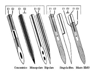
A motor unit (MU) includes a motor neuron, its axon, and the innervated muscle fibers. MU activity can be recorded using various electrode types: - **Single Fiber Electrode (SF-EMG):** Records one or two muscle fibers with a recording area of ~300 μm. Ideal for high-precision applications. Macro-EMG Needle Electrode:** Covers a large area (~15 mm) to record entire motor units. Concentric Electrode:** Balances recording precision and noise reduction with a 0.07 mm² area. - **Monopolar Electrode:** Larger recording area (~0.5-0.8 mm²), suitable for broader applications. MUPs represent the summation of action potentials from fibers within an MU. Key parameters include: Duration: Reflects MU size and fiber diameter. Amplitude:Indicates the number and proximity of fibers. Jiggle: Variability in MUP shape, indicative of reinnervation or pathology.
MUPs are evaluated using both manual and computerized methods: Template Matching: Identifies MUPs by comparing shapes.Signal Decomposition:Separates overlapping signals for detailed analysis.
The interference pattern reflects muscle contraction, analyzed through recruitment and frequency modulation mechanisms: - **Recruitment:** Progressive activation of MUs, starting with smaller, fatigue-resistant units.Frequency Modulation:Increase in MU firing frequency correlates with contraction strength.
Advanced methods like **Cloud analysis** plot IP parameters to distinguish myopathic from neuropathic conditions. In myopathy, points cluster below the "normal cloud," whereas in neuropathy, they fall above.
EMG provides invaluable insights into neuromuscular disorders: In 'myopathy', IP activity increases rapidly with reduced strength, reflecting smaller muscle fibers. In 'neuropathy', fewer MUs are activated, but reinnervation may increase MUP amplitude.
Conclusion: Clinical electromyography, with its advanced techniques and objective methodologies, remains an indispensable tool in neurology. Innovations like multiMUP and Cloud analysis enhance diagnostic precision, offering significant contributions to patient care.
To read the full text of this chapter, log in or request an account
A Google Account is needeed to request a Member Account
