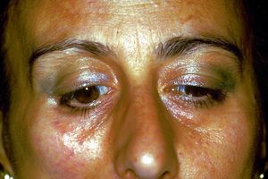
1° Klinischer Fall: Hemimastikatorischer Spasmus
In den vorangegangenen Kapiteln haben wir die diagnostische Komplexität in der Medizin betrachtet, wobei wir insbesondere einige Parameter berücksichtigt haben, wie die verborgenen Variablen, die uns nur die Entwicklung des Grundwissens im Laufe der Zeit ermöglichen wird, und die Schwierigkeiten bei der Entschlüsselung des verschlüsselten Signals, das das Zentralnervensystem in Form von verbaler Sprache nach außen sendet.
Nicht zuletzt die Einschränkungen durch eine deterministische Denkweise, die das Wissen in Fachkontexten reduziert, indem es seine diagnostische Kapazität einschränkt. Eine alternative Denkweise zur Deterministik, die in erster Linie eine Logik der Fuzzy-Sprache und den wichtigen Beitrag der Quantenwahrscheinlichkeit berücksichtigt, würde es uns ermöglichen, den wissenschaftlich-klinischen Horizont zu erweitern und in eine mesoskopische Realität einzutauchen.
In diesem Kapitel werden wir uns daher mit der Diagnose unserer Mary Poppins nach dem wissenschaftlich-klinischen Prozess befassen, die Schwierigkeiten der Implementierung und den Mehrwert des verschlüsselten Codes bewerten, der in der hepatischen Übertragung identifiziert wurde.
Einführung
Ausgehend von dem, was in den vorangegangenen Kapiteln von der „Einführung“ bis zu den Kapiteln „Logik der Medizinersprache“ dargelegt wurde, sahen wir uns über die Komplexität der Argumente und die Unbestimmtheit der verbalen Sprache hinaus mit einem Dilemma konfrontiert, dem des Kontexts die der Patient überwiesen wird, und in diesen Fällen scheint es für unsere arme Mary Poppins den zahnmedizinischen Kontext zu dominieren, angesichts der positiven Behauptungen, die aus den klinischen und Labortests hervorgehen, die an dem Patienten durchgeführt wurden.
(es scheint anscheinend aber ......)
Der klinische Fall unserer armen Mary Poppins zeigt die ganze physiopathologische und klinische Komplexität, aber vor allem ein Phänomen der Überschneidung von Aussagen, Aussagen und logischen Sätzen im zahnärztlichen und neurologischen Kontext, in dem ein Kontext Kompatibilität und Kohärenz erhält, während der andere Inkohärenz erhält.
Grundsätzlich angesichts der Kompatibilität und Konsistenz des Satzes (dentaler Kontext) mit den aus der klinischen Testberichterstattung abgeleiteten Behauptungen , sagen konsequent, dass orofaziale Schmerzen durch eine temporomandibuläre Störung verursacht werden, die inkompatibel werden könnten, wenn eine andere Reihe klinischer Aussagen auftritt stimmten mit dem Satz überein (neurologischen Kontext) und kontextuell gegenüber, aus diagnostischer Sicht, zu
Daher würde es eine Quelle für logistische Konflikte zwischen den beiden Fachkontexten mit unvermeidlicher diagnostischer Verzögerung geben, die auch den Entschlüsselungsprozess des Signals in der Maschinensprache des zentralen Nervensystems (ZNS) verunreinigen würde.
Was den Zugangsschlüssel zum Entschlüsseln des verschlüsselten Codes in Maschinensprache (SNC) bestimmt und es uns ermöglicht, eine Demarkationslinie abzufangen, werden unwiderlegbare klinische und / oder Labordaten genannt , was die Konsistenz einer Behauptung bestätigt oder ausschließt als die andere. Es ist an dieser Stelle wesentlich, a Satz (Abgrenzungssatz) des Typs wird sein:
Hat Mary Poppins eine neuromotorische Störung oder eine temporomandibuläre Störung?
Wir erklären diesen Schritt im Detail:
Denken Sie daran, dass da 'Die Logik der klassischen Sprache' dies zeigt:
- Eine Menge von sätze und a anzahl anderer aussagen, die sie logisch kompatibel sind, wenn, und nur wenn, die Vereinigung zwischen ihnen es ist konsequent
- Eine Menge von sätze und a anzahl anderer aussagen, die sie logisch inkompatibel sind, wenn, und nur wenn, die Vereinigung zwischen ihnen es ist inkonsequent
Bedeutung von Kontexten
Nun, für den zahnmedizinischen Kontext haben wir die folgenden Sätze und Behauptungen, denen wir einen numerischen Wert geben, um die Behandlung zu erleichtern, und das ist Wo zeigt "Normalität" und „Anormalität“ und damit Positivität des Berichts:
Positiver radiologischer Bericht des Kiefergelenks in Abbildung 2, Anomalie, Positivität des Berichts
Positiver CT-Bericht des Kiefergelenks in Abbildung 3, Anomalie, Positivität des Berichts
Positiver axiographischer Bericht der Kondylenspuren in Abbildung 4, Anomalie, Positivität des Berichts
Asymmetrisches EMG-Interferenzmuster in Abbildung 5, Anomalie, Positivität des Berichts
Der Satz (Zahnkontext) mit einer Zahl von anderen logisch kompatibel Behauptungen bestimmen die Vereinigung und Kohärenz zwischen ihnen und werden mit einem mathematischen Formalismus dargestellt, um die diagnostische Dialektik wie folgt zu erleichtern:
die den Durchschnitt der gemeldeten Einzelaussagen darstellt. Der Durchschnitt wurde entwickelt, weil es oft vorkommen kann, dass einige Tests negative Antworten geben, auf die der Wert gegeben werden muss
Das Ergebnis ist in diesem Fall und leitet zugleich die zusammenhängende Aussage des Satzes ab in dem argumentiert wird, dass die Symptomatologie der Patientin Mary Poppins durch das Vorhandensein eines DTMs bestimmt wird.
(und genau hier kollidieren die Kontexte)
Im neurologischen Kontext werden wir daher die folgenden Sätze und Behauptungen haben, denen wir einen Zahlenwert geben, um die Behandlung zu erleichtern, und das ist wo zeigt 'Normalität' an und „Anormalität“ und damit Positivität des Berichts:
Fehlen des Kieferrucks in den Abbildungen 6, Anomalie, Positivität des Berichts
Fehlen der stillen Masseterine-Periode in Abbildung 7, Anomalie, Positivität des Berichts
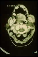
Hypertrophie des rechten Masseters im CT in Abbildung 8, Anomalie, Positivität des Berichts
Jetzt folglich auch Satz mit einer Nummer anderer logisch kompatibler Aussagen bestimmen die Vereinigung und Kohärenz zwischen ihnen und die formale Darstellung ähnelt der des zahnmedizinischen Kontexts:
mit kontextuell kohärenter Satzbejahung in dem argumentiert wird, dass die Symptomatologie der Patientin Mary Poppins eine neuromotorische Ursache hat und die klinischen Folgen eines Kau-Okklusionstyps nichts anderes als eine Folge der neuropathischen Schädigung sind.
Demarkator der Kohärenz
Der ist ein repräsentatives klinisches spezifisches Gewicht, komplex zu erforschen und zu verfeinern, weil es von Disziplin zu Disziplin und für Pathologien unterschiedlich ist, unerlässlich, um logische Behauptungen nicht kollidieren zu lassen und in den Diagnoseverfahren und wesentlich, um die Entschlüsselung des logischen Kommunikationscodes zu initialisieren. Grundsätzlich können Sie damit die Konsistenz einer Vereinigung bestätigen in Bezug auf einen anderenr und umgekehrt, wobei der Schwere der Aussagen und des Berichts im entsprechenden Kontext größeres Gewicht beigemessen wird.
Manchmal sieht sich der Arzt mit einer Reihe positiver Berichte konfrontiert, denen er gebührendes Gewicht beimessen muss. Er muss die Positivität eines Berichts berücksichtigen, der zum Beispiel hervorhebt, dass eine osteoartikuläre Remodellierung des Kiefergelenks nicht das gleiche Gewicht haben kann wie die Positivität eines bestätigenden Berichts eine Latenzverzögerung eines Trigeminusreflexes.
Das Gewicht der Abgrenzung , Daher gibt es den ernsthaftesten Behauptungen mehr Bedeutung in dem klinischen Kontext, aus dem sie stammen, und daher jenseits der Positivität der or Behauptungen, die ohnehin immer überprüft und respektiert werden, müssen diese mit a multipliziert werden wo zeigt 'Niedriger Schweregrad' und während „Hoher Schweregrad“.
Im neurologischen Kontext betrifft das Vorhandensein von Verzögerungen und/oder Latenz, das Fehlen des Kieferrucks, die Dauer und/oder das Fehlen der Schweigeperiode der Masseterine sowie andere Parameter, die im Moment nicht beschrieben werden müssen. Eine Verzögerung größer als der normative Wert oder das Fehlen der oben genannten Tests wird als schwerwiegendes und absolutes spezifisches Gewicht angesehen, das einer dominanten Wertigkeit in Bezug auf den anderen klinischen Kontext entspricht.
Zur Erinnerung haben wir daher:
wo
Durchschnitt der Wertigkeit klinischer Aussagen im zahnmedizinischen Kontext und damit
Durchschnitt der Wertigkeit klinischer Aussagen im neurologischen Kontext und damit
geringer Schweregrad Leistung des zahnärztlichen Kontextes Berichterstattung über einen hohen Schweregrad des neurologischen Kontextswo der 'Kohärenzmarker'' definiert den Diagnosepfad wie folgt
Sobald wir die unzähligen positiv gemeldeten normativen Daten weggespült haben, die dank des Kohärenzmarkers Konflikte zwischen Kontexten erzeugen wir haben ein viel klareres und lineareres Bild, um die Analyse der Funktionalität des zentralen Nervensystems zu vertiefen. Folglich können wir uns darauf konzentrieren, die Tests abzufangen, die zum Entschlüsseln des Maschinensprachcodes erforderlich sind, den der SNC in verbale Sprache umgewandelt aussendet.
Ephaptic transmission
With a little effort and patience on the part of passionate readers who have followed the entire logical path, sometimes apparently off topic, we have reached a clinical picture in which the code to be decrypted is inherent in neuromotor damage. Consequently, the access keys to the code, the one that figuratively corresponds to the exact decryption algorithm, would correspond to the right choice of the neuromotor damage detector test.
This is a point where it is essential to infer the clinician's intuition because finding the right algorithm to decrypt the code, after the aforementioned filter steps that have been described to wash away the interference of assertions, means at least choosing the right dictionary, for example, that of the image (MR of the brain rather than the neck); acoustic and vestibular evoked potentials (if a vestibular scwannoma is suspected) or trigeminal electrophysiological study (if more than one trigeminal involvement is suspected).
In this clinical iter that we have presented, the choice of the clinician to follow the electrophysiological trigeminal roadmap from which the positivity of the assertions have already been derived, therefore, having already defined a picture of serious anomaly of absence of the jaw jerk and of the silent period masseterino on the right side of the patient will have to understand if the damage is intracranial or extracranial.
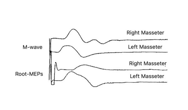
To do this, the clinician uses an electrical stimulation test of the masseter nerve in infratemporal fossa called on the masseter muscle with simultaneous recording of the heteronymous on the temporal muscle[1] and a bilateral transcranial electrical stimulation of the trigeminal motor roots called, precisely, [2]
M-wave
The right masseter nerve was electrically stimulated in the infratemporal fossa (see clinical procedure chapter: Encrypted code: Ephaptic transmission) with a technique similar to that described by Macaluso & De Laat (1995).[3] Square cathode pulses (0.1 ms) generated by an electrical stimulator (Neuropack X1, Nihon Kohden Corporation, Tokyo, Japan) were delivered through a Teflon-coated monopolar needle electrode (TECA 902-DMG25, 53534) with a tip non-isolated (diameter 0.36 mm; area 0.28 mm2) inserted 1.5 cm through the skin below the zygomatic arch and anterior to the temporomandibular joint in the infratemporal fossa with electrical shocks of 0.5 - 5 mA, and 0.1 ms. The anode was a surface non-polarizable Ag-AgCl disc electrode (OD 9.0 mm) positioned over the ipsilateral ear lobe. Electrical stimulation of the masseter nerve never produced pain, and the subjects only perceived muscle contraction. The correct position of the stimulation electrodes was monitored throughout the experimental session by checking online the size of the M wave in the masseter muscle. The signals were recorded by placing surface electrodes on the masseter and temporal muscles and filtered at 10-2000Hz and by concentric needle electrodes inserted into the anterior temporal muscle.
bRoot-MEPs
The trigeminal root was stimulated transcrally through high voltage, low impedance through an electrical stimulator (Neuropack X1, Nihon Kohden Corporation, Tokyo, Japan)) with the anode electrode positioned at the apex and the cathode approximately 10 cm laterally from the apex along a line vertex acoustic meatus. The electric field is believed to excite the trigeminal motor nerve fibers via the trancranial route, near their exit from the skull.[2][4] Also in this case, the response in the right masseter was markedly delayed (3.5 ms on the right side 2 ms on the left and dispersed. amplitude of the M-wave.
Having highlighted, through the execution of the and test, a delay in the conduction speed of the trigeminal nerve fibers generates the suspicion that it is a focal demyelination. This indicates that the problem is to be referred to the nervous component rather than to the muscular one, therefore, our attention should focus on the type of focal demyelination, extent of damage and presumably localization of the damage. The differential diagnosis at this point focuses on the type and area of the demyelinating damage, for example, if it is a damage exclusively referred to the masseterine motor nerve or the motor nerve of the temporal muscle is also involved, important for treatment with botulinum endotoxin. To resolve this doubt it is necessary to evoke a heteronymous response from recording on the temporal muscle.
H-wave
The arrangement is similar to that previously described with regard to the with the variant that the temporal muscle is recorded simultaneously with the stimulation of the masseterine nerve in the intratemporal fossa by a bipolar needle electrode. The stimulation must be gradually adapted in order to evoke both a from the masseter that a heteronymous from the temporal muscle.
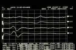
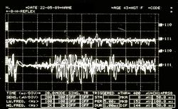
In our patient Mary Poppins, unfortunately, the neuromotor situation is quite complex and serious because to the infratemporal stimulation of the masseteral nerve we have a direct evoked response on the muscle ipsilateral to the stimulation (figure 10; ) and at the same time not a heteronymous response (figure 10; ) on the temporal muscle but rather a tonic asynchronous activity (Figure )
This neurophysiopathological phenomenon is called 'Ephaptic transmission' and allows us to confirm the presence of demyelinating lesions also of the motor nerve of the temporal muscle.
As previously mentioned and in order not to burden the topic, the detailed description of the 'Hephaptic' phenomenon will be treated in the chapter 'Encrypted code: Ephaptic transmission' in order to better understand the physiological responses of heteronymous by pathological ones.

Conclusions
Following this step by step path we have demonstrated a peripheral motor nerve injury as originally proposed by Kaufman.[5] Conduction studies have shown a slowing of conduction in the extracranial course of masticatory nerve fibers without a reduction in the amplitude of and obviously EMG signs of chronic denervation. The temporal muscle biopsy appeared histologically normal.
The absence of the jaw jerk on the diseased side indicates damage to the large diameter afferent fibers () from the neuromuscular spindles. A lesion of few afferents could easily abolish the jaw jerk.[6]
Muscle nerve damage would not be explained only because patients with Hemimasticatory Spasm (HMS) do not have sensory disturbances but also because they often only have spasms in one or two levator mandibular muscles. These observations argue against damage to the motor root or to the intracranial portion of the mandibular nerve where the motor bundles are closely grouped,[7] favoring damage to the individual muscle nerves that pass through the infratemporal fossa.
The mechanism of involvement of facial paroxysmal involuntary activity has been discussed by Kaufnan[5] and by Thompson and Carroll[8] who emphasized the close similarity between hemimasticatory and hemifacial spasm.
In EMG, these prolonged spasms fit perfectly into the description of cramps, that is, discharges of irregular motor units that progressively increase, leading to the recruitment of much of the muscle of synchronous discharges at speeds of 40 to 60 Hz.[9] Common to hemifacial spasm and cramps however, ectopic EMG activities can also be detected.
This could be responsible for the high frequency of EMG discharges at a frequency of 100-200 Hz and the synchronization of the entire muscle or multiple muscles, and post-activity. The synchronization could be explained by the lateral spread of discharges from adjacent nerve fibers,[10][11] generating a local re-excitation circuit. Posthumous EMG activity consists of paroxysmal discharges that may follow a voluntary orthodromic contraction or antidromic impulses,[12][13] and is attributed to self-excitation of the same axons after the passage of an impulse.
In our patient Mary Poppins we observed a synchronization of the whole or a large part of the muscle involved in the spasm (fig 10, EMG); the self-excitation is evidenced by the recording of the evoked discharges following the response of the stimulation of the chewing nerves (Fig. 10, E). These results support the hypothesis that spontaneous activity 'arises' in a demyelinated peripheral nerve, a phenomenon called hepaptic.[13]
In conclusion, the patient was affected by 'Hemimasticatory Spasm' mainly focused on the right masseter muscle but with indirect diffusion of the phenomenon to the right temporal muscle probably due to hepaptic activity due to the demyelination of the masticatory motor nerves in the infratemporal fossa. Botulinum endotoxin therapy was started immediately with total regression of the disease 10 years later.
(we will try to describe it in the chapter 'Crypted code: Ephaptic transmission' at the end of the section 'Hemasticatory spasm')
- ↑ Cruccu G, Truini A, Priori A, «Excitability of the human trigeminal motoneuronal pool and interactions with other brainstem reflex pathways», in J Physiol, The Physiological Society, 2001».
PMID:11230527 - PMCID:PMC2278464
DOI:10.1111/j.1469-7793.2001.0559i.x - ↑ 2.0 2.1 Frisardi G, «The use of transcranial stimulation in the fabrication of an occlusal splint», in J Prosthet Dent, Mosby, Inc - Elsevier, 1992».
PMID:1501190
DOI:10.1016/0022-3913(92)90345-b - ↑ Macaluso G, De Laat A. H-reflexes in masseter and temporalis muscle in man. Experimental Brain Research. 1995;107:315–320. [PubMed] [Google Scholar] [Ref list]
- ↑ G Frisardi, P Ravazzani, G Tognola, F Grandori. Electric versus magnetic transcranial stimulation of the trigeminal system in healthy subjects. Clinical applications in gnathology. J Oral Rehab.1997 Dec;24(12):920-8.doi: 10.1046/j.1365-2842.1997.00577.x.
- ↑ 5.0 5.1 Kaufman MD. Masticatory spasm in facial hemiatrophy. Ann Neurol 1980;7:585-7.
- ↑ Cruccu G, Inghilleri M, Fraioli B, Guidetti B, et al. Neurophysiologic assessment of trigeminal function after surgery for trigeminal neuralgia. Neurology 1987; 37:631-8.
- ↑ Pennisi E, Cruccu G, Manfredi M, Palladini G. Histometric study of myelinated fibers in the human trigeminal nerve. J Neurol Sci 1991;105:22-8.
- ↑ Thompson PD, Carroll WM. Hemimasticatory spasm: a peripheral paroxysmal cranial neuropathy? J Neurol NeurosurgPsychiatry 1983;46:274-6.
- ↑ Kimura J. Electrodiagnosis in diseases of nerve and muscle: principles and practice, 2nd edn. Philadelphia: FA Davis 1989.
- ↑ Nielsen VK. Pathophysiology of hemifacial spasm: II. Lateral spread of the supraorbital nerve reflex. Neurology 1984;34:427-31.
- ↑ Thompson PD. Stiff people. In Fahn S, Marsden CD, eds. Movement disorders 3. London: Butterworths, 1993: 367-99.
- ↑ Auger RG. Hemnifacial spasm: clinical and electrophysio- logic observations. Neurology 1979;29: 1261-72.
- ↑ 13.0 13.1 Nielsen VK. Pathophysiology of hemifacial spasm: I. Ephaptic transmission and ectopic excitation. Neurology 1984;34:418-26.
particularly focusing on the field of the neurophysiology of the masticatory system


![{\displaystyle \delta _{n}=[0|1]}](https://wikimedia.org/api/rest_v1/media/math/render/svg/cd35df3912a48e5b0b70d9cd5b2e1bee432c3272)


![{\displaystyle \gamma _{n}=[0|1]}](https://wikimedia.org/api/rest_v1/media/math/render/svg/2f85d0ed73fa3e7903f0321e48668467c1277f4e)





![{\displaystyle \tau =[0|1]}](https://wikimedia.org/api/rest_v1/media/math/render/svg/fbdc534cec0dcf1f070a551e40611eb83e8aca25)


