Encrypted code: Bilateral Motor Evoked Potentials of trigeminal root
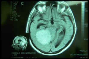
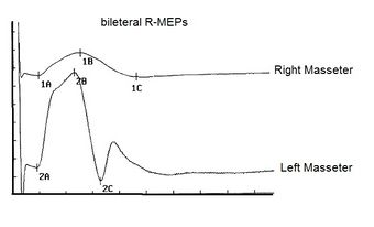
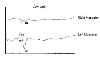
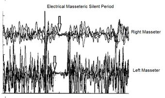
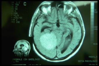
In this last chapter referring to the correlation between dental malocclusion and postural disorders we can perceive how the axioms sometimes hastily generated from a limited basic knowledge in the field of trigeminal neurophysiopathology can cause serious damage from a diagnostic point of view. The patient 'Balancer' presented in the previous chapters had been suffering for more than 10 years from a meningioma at the base of the skull which, due to its progressively growing volume, compressed and simultaneously stretched the sensory and motor fibers of the trigeminal system as well as damaging the midbrain centers adjacent to the tumor. The term used by the patient in reporting the disturbance in verbal language, was 'chewing difficulty' but in machine language it should have been decrypted in 'lack of the masticatory stereognosic effect' due to proprioceptive sensory deficit. The absence of the jaw jerk on the right side, in fact, demonstrates the real extent of the damage. The bRoot-MEPs confirm the organic damage and the latency asymmetry of the electrical silent period concludes the clinical diagnosis. MRI shows the severe neurological complication of brainstem displacement. It should be noted that a meningioma of this size increases in volume until it reaches, as in our case, a diameter of 8 cm, with a ten-year time latency. The question is the following: in the early stages of the volumetric increase where only masticatory-type disturbances reported to dental colleagues were appreciable, would it have been possible to make an electrophysiological diagnosis of organic trigeminal damage? Of course yes, because, perhaps we would not have witnessed an absence of reflexes both in latency and in amplitude but we would certainly have noticed a significant asymmetry and a latency delay of the silent period as well as an initial amplitude anomaly in the bRoot-MEPs. In conclusion, the correlation between the vestibular and trigeminal system, although present from an anatomical and neurophysiopathological point of view, should not be considered by masticatory rehabilitation clinical procedures because an error in the differential diagnosis is too high and dangerous.
Introduction
In the introductory chapter concerning the 3rd clinical case affected by meningioma in which the masticatory difficulty reported by the patient 'Balancer' had been correlated to a prosthetic rehabilitation discrepancy, we have already arrived at a first diagnostic filter considering the neurological assertion to be valid than the dental one. Consequently, one can concentrate on intercepting the tests necessary to decrypt the machine language code that the CNS sends out converted into verbal language. Apparently this verbal language would address the case in a Postural disorder related to a dental malocclusion due to incongruous prosthetic rehabilitation. If, on the one hand, there may be an asymmetry of the interferential EMG of the masseters due to a prosthetic occlusal imbalance, on the other, such an evident asymmetry of the jaw jerk and the silent period cannot be justified. For this reason, it is essential to continue with the Masticationpedia diagnostic model in order to arrive at an exact and rapid conclusive diagnosis. We therefore begin with the 'Cognitive Neural Network' which responds, as we know by now, with a sequence of scientific-clinical data, always taking into consideration the importance of the 'initiation' step which in this case has been set in 'Gait'.
Gait (45.300), motor evoked potentials (231),reflex (36), jaw(1) 'Cerebellar ataxia, neuropathy, vestibular areflexia syndrome (CANVAS) with chronic cough and preserved muscle stretch reflexes: evidence for selective sparing of afferent Ia fibres'
Diagnostic sequence
1st Step: CNN Sequence
- Coherence Demarcator: As we have previously described for the other clinical cases, the first step is an initialization command of the Cognitive Neural Network 'CNN' which derives, in fact, from a previous cognitive elaboration on the assertions in the dental and neurological context in which the ' Demarcator of Consistency' gave a prevailing weight. The dental context has already been eliminated from the Consistency Demarcator. From what emerges from the neurological statements, the 'State' of the Trigeminal Nervous System' appears to be strongly damaged. The initialization command will therefore be 'Gait'.
- 1st loop open: The first result of the 'CNN' for the keyword 'Gait' returns 45,300 results, obviously, too vast to be exhaustive but having ascertained in the context analysis phase a serious trigeminal electrophysiological situation with marked structural anomalies and functional, we can safely enter a correlated key such as ' Motor Evoked Potentials' without specifying the trigeminal district which could encroach on the identified set.
- 2st loop open: To this second query 'Motor Evoked Potentials' the database replies with 231 results that are still too vast as an answer and therefore we are looking for a key more similar to the clinical case presented. Since the most anomalous results in the neurological context have emerged from the latency and amplitude alterations of the trigeminal reflexes, an appropriate access key could be, in fact, 'Reflex' also without specifying 'trigeminal' for the same anticipated reasons.
- 3st loop open: To the 'Reflex'request, the response was 36 results which narrowed the field of analysis for the diagnosis of our patient 'Balancer'. Only at this level of the CNN can one attempt to close the loop with a more specific request such as 'Jaw'. In this way we have not lost contact with the whole considered and we have remained in the field of electrophysiology.
- 4st loop open: The request 'jaw' in fact in the 36 results it is possible to intercept an article in which some electrophysiological parameters are reported which correspond to a clinical situation of cerebellar ataxia, a pathology in which postural and gait instability is a clinical sign impressive and important.
2st Step: CNN analysis
The 'CNN' loop closure analy of course is based on the terminal article which basically describe five patients with cerebellar ataxia, neuropathy and vestibular areflexia syndrome (CANVAS) with chronic cough and lower limb muscle stretch reflexes preserve yourself.[1] In particular, somatosensory evoked potentials were absent or severely attenuated. Biceps and hamstring T-reflex recordings were normal, while the masseter reflex was absent or attenuated.
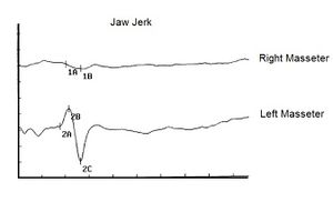
The first observation to be made is that the patients were suffering from chronic spasmodic cough and the second observation was the preservation of the tendon reflexes of the lower limbs. In our patient 'Balancer', on the other hand, there was a total absence of the mandibular tendon reflex[2] () so that the neurological damage was very evident at the trigeminal midbrain level. (Figure 1) The multifunctional contribution of the midbrain synaptic circuitry by the proprioceptive nerve endings ( e ) are of primary importance both for posture and for cervico-oculomotor reflexes. A very interesting article by Yongmei Chen et al.[3] showed, through markers, how neurons afferent to the trigeminal mesencephalic nucleus (Vme) from the jaw muscles project to the oculomotor nuclei (III/IV) and their premotor neurons in the interstitial nucleus of Cajal (INC), a well-known pre-oculomotor center that vertically manipulates torsional eye movements.
The conceptual conclusion of the authors was that the Vme proprioceptive neurons of the masticatory projecting muscles at III/IV and INC would detect spindle activity to spatial changes of the jaw conditioned by the force of gravity and/or by the connection between the mandible during rotation of the head. Thus, the convergent innervation of Vme and MVN neurons on the oculomotor and pre-oculomotor nuclei would be a neuroanatomical substrate for the interaction of masticatory proprioception with vestibulo-ocular signals on the oculomotor system during vertical-torsional VOR. The contribution of this article obviously allows us to consider a correlation between the trigeminal system, posture and gait, therefore, the abnormal asymmetry of the jaw jerk could be related to a postural disorder of our patient 'Balancer'
But there is also to consider the article of Jon Infante [1] highlighted that all five patients were in the seventh decade of age, their gait imbalance having started in the fifth decade. In four patients, cough preceded gait imbalance between 15 and 29 years of age; the cough was spasmodic and triggered by variable factors. In addition, vestibular function tests showed bilateral impairment of the vestibulo-ocular reflex.
The presence of chronic spasmodic cough, in this study, for over 10 years even before balance and postural disturbances showed up, suggest that machine language, as repeatedly exposed in the specific chapters of Masticationpedia, is a reality not to be overlooked because the ambiguous and vague verbal language covers the information of the encrypted code. As we will demonstrate in the course of the discussion, the analog of the spasmodic cough symptom of the patients reported by Jon Infante's article could be the repeated reporting to the dental colleagues of the chewing difficulty by our patient 'Balancer'. (Figure 2) This parameter has not been considered as an 'encrypted code' but only an element of verbal language logic and therefore vague and ambiguous. The continuous remaking of the prosthetic rehabilitation dampened even more the information of the machine language until the temporal and spatial addition of the organic damage was transformed[1] into a macroscopic symptom of vertigo and loss of balance in walking
Jon Infante's article[1] concludes with a striking statement, i.e. that spasmodic cough can be an integral part of the clinical picture in CANVAS, anticipating the appearance of postural imbalance by several decades and that sparing of muscle spindle afferents (Ia fibers) is probably the pathophysiological basis of normoreflexia.
(........is the neurological damage functional or organic?)
Decryption process
L'iter diagnostico seguito ormai di routine secondo il modello Masticationpedia ci ha consentito di eliminare, attraverso il demarcatore di coerenza , il contesto odontoiatrico per seguire quello neurologico e di approfondire attraverso lo 'Cognitive Neural Network' (CNN) le possibili correlazioni tra sintomi e anomalie neurofisiologiche nei pazienti con disturbi posturali, di deambulazione dovute a patologie funzionali ( labirintiti, neuropatie ecc.) da quelle organiche ( tumori, demielinizzazioni ecc.). Ora bisogna cercare di decriptare il messaggio criptato del linguaggio macchina del Sistema Nervoso Trigeminale.
Codice criptato
Come anticipato, in primis abbiamo potuto evidenziare una grave assenza del jaw jerk riportata come Anormalità, positività del referto ( Figura 1) ma come è stato già menzionato nel capitolo ' Conclusioni sullo status quo nella logica del linguaggio medico riguardo al sistema masticatorio' anche nel paziente trattato con chirurgia ortognatica abbiamo potuto rilevare una assenza del jaw jerk (figura 2) ripristinato subito dopo riabilitazione neurognatologica evocata. Nel nostro caso 'Balancer' abbiamo un disturbo oggettivo in più quello della difficoltà masticatoria ed insieme al disturbo posturale erano presenti nistagno e vertigini.
Un ulteriore asserzione a favore del danno organico è stato anche la presenza di un ritardo di latenza sul lato destro del periodo silente elettrico, Anormalità, positività del referto, che depone per un rallentamento della velocità di conduzione nervosa. In questo istanza non possiamo dire se il ritardo sia riferito ad un danno delle fibre sensitive o di quelle motorie fintanto che non si approccia allo studio della conduzione nervosa delle radici trigeminali motorie.
Potenziali Evocati Motori della radice trigeminale
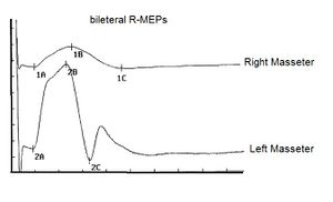
Nel nostro laboratorio di neurofisiologia masticatoria abbiamo messo a punto una tecnica di elettrostimolazione trasncraniale elettrica delle due radici trigeminali in simultanea e sincronizzate con lo stimolo elettrico. Nei vari capitoli già pubblicati sono riportati alcune informazioni tecniche sul metodo ma ci ripromettiamo di esporli in modo esaustivo nella sezione 'Scienza straordinaria'. In questo contesto possiamo solo considerare e confermare una asimmetria notevole di ampiezza delle risposte evocate motorie come mostrato in figura 3. I markers 1A e 2A indicano la latenza chea differenza del periodo silente è simmetrica e questo dato conferma il danno strutturale delle fibre sensitive comprese quelle propriocettive dai muscoli masticatori. Possiamo a questo punto non solo confermare e giustificare la scelta neurologica del contesto diagnostico ma anche concludere con una pre diagnosi di danno neurologico cerebellare con coinvolgimento dell'asse mesencefalico trigeminale.
La Risonanza Magnetica dell'encefalo, purtroppo, risolve i nostri dubbi con una refertazione di lesione espansiva solida con effetto massa nell'emisfero destro, non è chiaro se abbia un’origine sopra o sotto tentoriale, sicuramente determina fenomeni compressivi sul mesencefalo - tronco e sul quarto ventricolo con dilatazione delle cisterne e dei ventricoli a monte. Che si tratti di un meningioma è molto probabile perché non ha edema perilesionale.
Final considerations
Riassumiamo il percorso clinico diagnostico seguendo il modello Masticationpedia perched si possa considerare quest'ultimo un fluido e dinamico iter per giungere a target diagnostico nel modo più rapido e dettagliato.
- Analisi dei contesti attraverso test di laboratorio odontoiatrici e neurologici
- Scelta dell'asserzione neurologica attraverso filtro da parte del Demarcatore di Coerenza
- Valutazione del caso clinico nel Cognitive Neural Network (CNN)
- Chiusura del loop del CNN con l'articolo di Jon Infante [1] e la considerazione del danno organico/funzionale delle strutture sensitivo motorie trigeminali del nostro paziente attraverso constatazione della asimmetria di ampiezza della 'bRoot-MEPs'. Nella galleria di immagini è stata ordinata la sequenza logica dei test eseguiti con ripristino della numerazione.
Figura 1c: Dopo aver verificato il livello di integrità dell'area mesencefali attraverso test che coinvolgono riflessi monosinaptici come il jaw jerk si alza il livello di complessità evocando il periodo silente elettrico che è un riflesso polisinaptico. Appare evidente il ritardo di latenza sul massetere destro
Conclusioni
Abbiamo ormai evidenze di correlazione organico funzionali sia dirette che indirette tra il sistema trigeminale ed il sistema vestibolare, basti pensare allo studio dei mVEMP (Vestibular Evoked Myogenic Potentials) ormai riconosciuto come un test solido e affidabile per valutare l'integrità funzionale della via del riflesso vestibolo-masseterico,[4] nelle manifestazioni cliniche con coinvolgimenti del sistema trigeminale e vestibolare come nei schwannomi[5][6] od in presenza di neurinomi dell'acustico[7] tanto quanto la relativa correlazione tra occlusione dentale e sistema vestibolare ma ciò non consente, visto la gravità dell'errore diagnostico che ne deriverebbe, di considerare quest'ultima condizione clinica un dato scientifico validato clinicamente. InAppendice un esempio di ciò che potrebbe accadere.
Quest'ultima asserzione, forse rischiosa perchè di controversione, si scontra sull'evidenza di un fatto eclatante più volte evidenziato nel corso della stesura dei capitoli di Masticationpedia quello del linguaggio macchina e del linguaggio verbale. Un linguaggio verbale vago ed ambiguo può generare convinzioni scientifiche a seconda di come viene proposto ma in sostanza copre ed occulta un linguaggio macchina molto più determinante e formale. Per esempio il linguaggio verbale ha dedotto che i disturbi posturali del paziente 'Balancer' fossero causati da una incongrua riabilitazione protesica mentre il linguaggio macchina segnalava un deficit neurosensoriale organico ( assenza del jaw jerk, ritardi in latenza del periodo silente, ampia riduzione di ampiezza della bRoot-MEPs.
La mancanza dell'informazione della velocità e della posizione mandibolare a livello mesencefalico, infatti, determina una incapacità di coscienza stereognosica in cui il paziente non riconosce la posizione spaziale e temporale della mandibola. La diminuzione del reclutamento delle unità motorie dei muscoli masticatori e non solo del massetere testato con una ovvia diminuzione della forza masticatoria è il risultato di questo danno organico e funzionale. Tutto ciò indica una compromissione del territorio propriocettivo ma non sappiamo se sono stati coinvolti anche le strutture sensitive. Il periodo silente elettrico mostra, invece, un grave ritardo in latenza con un rallentamento della velocità di conduzione nervosa. A concludere il quadro diagnostico neurofisiopatologico è la constatazione che la radice motoria trigeminale di destra ha perso gran parte delle proprie fibre motorie.
Da notare, però, che in una condizione clinica così grave assistiamo ad una diminuzione dell'ampiezza della bRoot-MEPs ma non una asimmetria di latenza. Questo risultato indica che c'è stato un danno diretto sulle fibre motorie trigeminali dalla compressione del tumore ( assonotmesi) ma non una demielinizzazione (neuroprassia) che avrebbe mostrato un ritardo di latenza sul massetere destro. E' bene descrivere brevemente la definizione di danno neurologico tenendo conto che il corrispettivo del nervo periferico spinale per il sistema nervoso trigeminale corrisponde alla radice trigeminale motoria.
La neurotmesi è causata dalla transezione di un nervo ed è il peggior grado di lesione del nervo periferico. Nella neurotmesi, l'intero nervo, compresi l'endoneurio, il perinevrio e l'epinevrio, è completamente reciso. La neurotmesi porta alla rottura dell'assone, della guaina mielinica e dei tessuti connettivi. La prognosi per il recupero spontaneo è infausta senza intervento chirurgico.[8] La lesione di quinto grado di Sunderland corrisponde alla definizione di neurotmesi nella classificazione di Seddon e rappresenta il più alto grado di lesione nervosa, con un difetto completo del nervo.
La neuroaprassia è una lesione nervosa comunemente indotta da demielinizzazione focale e/o ischemia ed è il tipo più lieve di lesione del nervo periferico. Nella neuroaprassia, la conduzione degli impulsi nervosi è bloccata nell'area lesa, la connessione motoria e sensoriale è persa, ma tutte le strutture morfologiche del moncone nervoso, inclusi l'endoneurio, il perinevrio e l'epinevrio, rimangono intatte.
L'assonotmesi è un tipo relativamente più grave di lesione del nervo periferico e di solito è causata da schiacciamento, stiramento o percussione. Nell'assonotmesi, l'epinevrio è intatto, mentre il perinevrio e l'endoneurio possono essere interrotti. L'assone viene separato dal soma e l'assone e la guaina mielinica vengono interrotti. La degenerazione walleriana si verifica nel moncone dell'assone distale al sito della lesione entro 24-36 ore dopo la lesione del nervo periferico.
Dopo la neurotmesi, le cellule di Schwann rispondono in modo adattivo all'interruzione assonale, passando da uno stato altamente mielinizzato a uno stato de-differenziato. Le cellule di Schwann de-differenziate fagocitano assoni e detriti di mielina e formano un percorso di rigenerazione per la crescita degli assoni. Inoltre, le cellule di Schwann attivate secernono un gruppo di citochine, tra cui il fattore di necrosi tumorale-alfa, l'interleuchina-1 alfa e il fattore inibitorio della leucemia, per reclutare i macrofagi e facilitare la digestione dei detriti. Le cellule di Schwann secernono anche un gruppo di fattori neurotrofici, tra cui il fattore di crescita nervoso, il fattore neurotrofico derivato dal cervello e il fattore neurotrofico derivato dalla linea cellulare gliale, per incoraggiare la sopravvivenza dei neuroni e l'allungamento degli assoni.[9][10][11]
L'elettromiografia con ago (EMG) è lo studio elettrodiagnostico più sensibile per la perdita dell'assone motorio e, con lesioni di grande gravità, compaiono risposte motorie di bassa ampiezza. Il decremento dell'ampiezza della risposta motoria inizia intorno ai giorni 2-3 ed è completo entro il giorno 6. Ciò riflette il fatto che la degenerazione della giunzione neuromuscolare precede la degenerazione degli assoni e le risposte motorie dipendono dalla trasmissione della giunzione neuromuscolare.[12]
Appendice
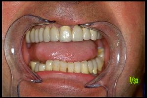
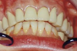
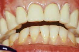
Paziente trattata da collega odontoiatrico per una riabilitazione protesica seguendo metodiche posturali che attraverso pedane dinamomentriche indicavano la migliore occlusione Centrica. La paziente giunse alla nostra osservazione presentando uno stato clinico di fasciolazione dei muscoli masseteri oltre, ovviamente, un discomfort occlusale e masticatorio. La posizione centrica finalizzata dal collega era, perciò, correlata con la migliore risposta dinamometria posturale ma purtroppo così non é risultato.
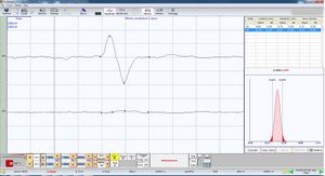
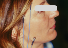
Osservando le figure 4,5 e 6 possiamo comprendere come si costruiscono assiomi che non rispondono ad un criterio scientifico convalidato tanto meno ad una realtà biologica Pensare che un fenomeno a distanza come la stabilità posturale dettata da una pedana dinamometria possa convalidare la scelta di una posizione occlusale spaziale raggiunta manualmente dall'operatore si infrange sulla constatazione che osservando lo stato di sistema trigeminale misurandolo attraverso un demand ( bRoot-MEPs) il sistema risponde con una propria posizione spaziale non dettata ne dalla correlazione posturologica ne tanto meno dalle manovre manuali dell'operatore bensì da una proprio risultato vettoriale neuromotorio.
(….dovrebbe esclusivamente essere considerata come campo esplorativo sperimentale per dedurre conoscenze maggiori sulla connettivitá neuronale.)
- ↑ 1.0 1.1 1.2 1.3 1.4 Jon Infante, Antonio García, Karla M Serrano-Cárdenas, Rocío González-Aguado, José Gazulla, Enrique M de Lucas, José Berciano. Cerebellar ataxia, neuropathy, vestibular areflexia syndrome (CANVAS) with chronic cough and preserved muscle stretch reflexes: evidence for selective sparing of afferent Ia fibres.J Neurol . 2018 Jun;265(6):1454-1462. doi: 10.1007/s00415-018-8872-1.Epub 2018 Apr 25.
- ↑ The history of examination of reflexes. Boes CJ.J Neurol. 2014 Dec;261(12):2264-74. doi: 10.1007/s00415-014-7326-7. Epub 2014 Apr 3.PMID: 24695995
- ↑ Chen Y, Gong X, Ibrahim SIA, Liang H, Zhang J.. Convergent innervations of mesencephalic trigeminal and vestibular nuclei neurons onto oculomotor and pre-oculomotor neurons-Tract tracing and triple labeling in rats. PLoS One. 2022 Nov 28;17(11):e0278205. doi: 10.1371/journal.pone.0278205. eCollection 2022.PMID: 36441755
- ↑ Sangu Srinivasan Vignesh, Niraj Kumar Singh, Krishna Rajalakshmi. Tone Burst Masseter Vestibular Evoked Myogenic Potentials: Normative Values and Test-Retest Reliability. J Am Acad Audiol. 2021 May;32(5):308-314. doi: 10.1055/s-0041-1728718.Epub 2021 Jun 1.
- ↑ Ashutosh Kumar, Sanjay Behari, Jayesh Sardhara, Prabhaker Mishra, Vivek Singh, Vandan Raiyani, Kamlesh Singh Bhaisora, Arun Kumar Srivastava . Quantitative assessment of brainstem distortion in vestibular schwannoma and its implication in occurrence of hydrocephalus.Br J Neurosurg . 2022 Dec;36(6):686-692. doi: 10.1080/02688697.2022.2047155.Epub 2022 Mar 7.
- ↑ Daniel Moualed, Jonathan Wong, Owen Thomas, Calvin Heal, Rukhtam Saqib, Cameron Choi, Simon Lloyd, Scott Rutherford, Emma Stapleton, Charlotte Hammerbeck-Ward, Omar Pathmanaban, Roger Laitt, Miriam Smith, Andrew Wallace, Mark Kellett, Gareth Evans, Andrew King, Simon Freeman. Prevalence and natural history of schwannomas in neurofibromatosis type 2 (NF2): the influence of pathogenic variants. Eur J Hum Genet. 2022 Apr;30(4):458-464. doi: 10.1038/s41431-021-01029-y.Epub 2022 Jan 24.
- ↑ Claudia Cassandro, Roberto Albera, Luca Debiasi, Andrea Albera, Ettore Cassandro, Alfonso Scarpa, Massimo Ralli. What factors influence treatment decision making in acoustic neuroma? Our experience on 103 cases. Int Tinnitus J. 2020 Nov 18;24(1):21-25.doi: 10.5935/0946-5448.20200004.
- ↑ Kaya Y, Sarikcioglu L. Sir Herbert Seddon (1903-1977) and his classification scheme for peripheral nerve injury. Childs Nerv Syst. 2015 Feb;31(2):177-80.
- ↑ Jessen KR, Mirsky R, Lloyd AC. Schwann Cells: Development and Role in Nerve Repair. Cold Spring Harb Perspect Biol. 2015 May 08;7(7):a020487.
- ↑ Madduri S, Gander B. Schwann cell delivery of neurotrophic factors for peripheral nerve regeneration. J Peripher Nerv Syst. 2010 Jun;15(2):93-103.
- ↑ Yi S, Zhang Y, Gu X, Huang L, Zhang K, Qian T, Gu X. Application of stem cells in peripheral nerve regeneration. Burns Trauma. 2020;8:tkaa002
- ↑ Ferrante MA. The Assessment and Management of Peripheral Nerve Trauma. Curr Treat Options Neurol. 2018 Jun 01;20(7):25.
