Intermittierende Gesichtskrämpfe als Zeichen eines rezidivierenden pleomorphen Adenoms
| Title | Intermittierende Gesichtskrämpfe als Zeichen eines rezidivierenden pleomorphen Adenoms |
| Authors | Rosalie A Machado · Sami P Moubayed · Azita Khorsandi · Juan C Hernandez-Prera · Mark L Urken |
| Source | Document |
| Date | 2017 |
| Journal | World J Clin Oncol |
| DOI | 10.5306/wjco.v8.i1.86 |
| PUBMED | https://pubmed.ncbi.nlm.nih.gov/35287260/ |
| PDF copy | |
| License | Template:CC BY-NC |
| This resource has been identified as a Free Scientific Resource, this is why Masticationpedia presents it here as a mean of gratitude toward the Authors, with appreciation for their choice of releasing it open to anyone's access | |
This is free scientific content. It has been released with a free license, this is why we can present it here now, for your convenience. Free knowledge, free access to scientific knowledge is a right of yours; it helps Science to grow, it helps you to have access to Science
This content was relased with a Template:CC BY-NC license.
You might perhaps wish to thank the Author/s
Intermittierende Gesichtskrämpfe als Anzeichen eines rezidivierenden pleomorphen Adenoms
Free resource by Rosalie A Machado · Sami P Moubayed · Azita Khorsandi · Juan C Hernandez-Prera · Mark L Urken
|
Abstrakt
Die enge anatomische Beziehung des Gesichtsnervs zum Parotisparenchym hat einen erheblichen Einfluss auf die auftretenden Anzeichen und Symptome sowie die Diagnose und Behandlung von Parotisneoplasien. Allerdings wurde unseres Wissens nach eine Hyperaktivität dieses Nervs, die sich als Gesichtskrampf äußert, nie als Anzeichen oder Symptom einer bösartigen Erkrankung der Ohrspeicheldrüse beschrieben. Wir berichten über einen Fall eines Karzinoms, das aus einem rezidivierenden pleomorphen Adenom der linken Ohrspeicheldrüse (d. h. einem Karzinom ex pleomorphem Adenom) entstand und mit hemifazialen Spasmen einherging. Wir skizzieren die Differentialdiagnose des Hemifacialispasmus sowie eine vorgeschlagene Pathophysiologie. Gesichtslähmungen, Lymphknotenvergrößerungen, Hautbeteiligung und Schmerzen wurden alle mit bösartigen Erkrankungen der Ohrspeicheldrüse in Verbindung gebracht. Bisher wurde nicht über die Entwicklung eines Gesichtsspasmus bei bösartigen Erkrankungen der Ohrspeicheldrüse berichtet. Die häufigsten Ursachen für hemifazialen Spasmus sind Gefäßkompression des ipsilateralen Gesichtsnervs im Kleinhirnbrückenwinkel (als primär oder idiopathisch bezeichnet) (62 %), erblich bedingt (2 %), sekundär zu Bell-Lähmung oder Gesichtsnervenverletzung (17 %). Hemifaziale Spasmen imitieren (psychogen, Tics, Dystonie, Myoklonus, Myokymie, Myorthythmie und hemimastikatorischer Spasmus) (17 %). Hemifacialer Spasmus wurde nicht im Zusammenhang mit einem bösartigen Parotistumor berichtet, muss aber bei der Differenzialdiagnose dieses vorliegenden Symptoms berücksichtigt werden.
Schlüsselwörter: Gesichtskrampf, pleomorphes Adenom, gutartiger gemischter Parotistumor, rekonstruktive Chirurgie, Speicheldrüsen
Kerntipp: Dieser Bericht stellt den ersten Fall eines hemifazialen Spasmus dar, der mit der Umwandlung eines rezidivierenden pleomorphen Adenoms in ein Karzinom ohne pleomorphes Adenom einhergeht. Die Ursache hemifazialer Spasmen wird diskutiert.
Gehe zu:
EINFÜHRUNG
Die enge anatomische Beziehung des Gesichtsnervs zur Ohrspeicheldrüse hat einen erheblichen Einfluss auf die Symptome/Anzeichen, die Diagnose und die Behandlung von Ohrspeicheldrüsenneoplasien[1]. Eine Beteiligung des Gesichtsnervs durch bösartige Erkrankungen der Ohrspeicheldrüse führt in der Regel zu einer teilweisen oder vollständigen Hemifazialen Lähmung[2]. Unseres Wissens wurde jedoch nicht über eine Hyperaktivität dieses Nervs, die sich als Gesichtskrampf äußert, als charakteristisches Merkmal eines bösartigen Parotistumors berichtet. Dennoch wurde in der Literatur zweimal über einen Gesichtsspasmus als charakteristisches Merkmal eines gutartigen Parotistumors berichtet[3]. Wir berichten über einen Fall eines Karzinoms, das bei einem rezidivierenden pleomorphen Adenom (d. h. Karzinom ex pleomorphem Adenom) auftrat und mit hemifazialen Spasmen einherging. Wir skizzieren die Differentialdiagnose des Hemifacialispasmus sowie eine vorgeschlagene Pathophysiologie.
Dies ist ein einzelner institutioneller Fallbericht in einem tertiären Überweisungskrankenhaus. Das Institutional Review Board war nicht verpflichtet, einen Fall an unserer Einrichtung zu melden.
Gehe zu:
FALLBERICHT
Eine 56-jährige Raucherin hatte in der Vorgeschichte ein pleomorphes Adenom in der linken Ohrspeicheldrüse, das im Alter von 18 Jahren mit einer oberflächlichen Parotidektomie behandelt wurde. Neunzehn Jahre nach dieser Operation stellte sich bei der Patientin ein multifokales Rezidiv vor. Es wurde eine chirurgische Untersuchung durchgeführt und es wurde festgestellt, dass der Tumor untrennbar mit dem Gesichtsnerv verbunden war. Zu diesem Zeitpunkt wurde die Resektion abgebrochen und der Gesichtsnerv nicht geopfert, und im Ohrspeicheldrüsenbett blieb eine schwere Erkrankung zurück. Der Patient unterzog sich einer externen Strahlentherapie und die Größe des Tumors blieb bei der Überwachung durch serielle Computertomographie (CT) und Magnetresonanztomographie (MRT) 10 Jahre lang stabil. Die Patientin war klinisch asymptomatisch, bis sie anfing, intermittierende ipsilaterale hemifaziale Krämpfe zu entwickeln, die spontan auftraten und alle Teile der linken Gesichtsmuskulatur betrafen, was sie dazu veranlasste, zur Untersuchung zurückzukehren.
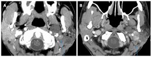
Wiederholte CT-Scans zeigten eine Vergrößerung des sich stark und gleichmäßig vergrößernden soliden Tumors ohne Bereiche mit Nekrose oder extrakapsulärer Ausdehnung mit Ausbreitung in das linke Foramen stylomastoideum, zusammen mit verdächtigen Veränderungen im vergrößerten (15 mm) linken Lymphknoten der Ebene IV (Abbildung (Abbildung 1A).1A ). Eine Feinnadelaspirationsbiopsie des Tumors ergab den Verdacht auf ein Karzinom ohne pleomorphes Adenom. Nach einer negativen systemischen Metastasenuntersuchung wurde der Patient für eine radikale Parotidektomie mit Tötung des Gesichtsnervs, ipsilateraler selektiver Halsdissektion (Level I-IV) und einem deepithelisierten anterolateralen freien Oberschenkellappen zur Volumenwiederherstellung in den Operationssaal gebracht um die Wundheilung zu verbessern. Der vertikale Abschnitt des Gesichtsnervs im Mastoid wurde freigelegt. Die Reparatur des primären Gesichtsnervs erfolgte durch eine Transplantation des Nervus suralis vom Hauptstamm auf den Schläfenast des Gesichtsnervs, eine Transplantation des Nervs auf den Masseter an den dominanten Mittelgesichtsästen des Gesichtsnervs sowie die Konstruktion einer oralen Kommissursuspension mit einer Fascia-lata-Schlinge .

Die abschließende chirurgische Pathologie bestätigte ein 5,2 cm großes pleomorphes Adenom mit einem multinodulären Wachstumsmuster. Gut umschriebene neoplastische Knötchen unterschiedlicher Größe waren in dicht fibrotischem Bindegewebe eingebettet (Abbildung (Abbildung 2).2). Im Narbengewebe zwischen den Knötchen waren auch Nervenbündel eingeklemmt, eine echte perineurale Invasion konnte jedoch nicht festgestellt werden. Innerhalb der Knötchen wurden zwei Herde eines frühen nicht-invasiven Karzinoms festgestellt. Innerhalb eines Knotens wurde ein 4 mm großer Herd maligner Zellen identifiziert, der von gutartigen Epithelelementen umgeben war. In einem separaten Knoten wurde außerdem eine intraduktale maligne neoplastische Proliferation mit einem intakten gutartigen myoepithelialen Zellrand festgestellt. Keiner der bösartigen neoplastischen Herde zeigte eine Invasion in angrenzendes fibroadiöses Gewebe und Nerven. Dreizehn Lymphknoten der Stufen II–V waren negativ hinsichtlich einer Tumorbeteiligung. Der Primärtumor wurde als rT4N0M0 eingestuft.
Die hemifazialen Krämpfe ließen nach der Operation nach und der Patient blieb nach 6 Monaten Nachuntersuchung krankheitsfrei. Der Gesichtstonus des Patienten ist wiederhergestellt, die dynamische Muskelaktivität ist jedoch noch nicht entwickelt.
Gehe zu:
DISCUSSION
Zbären et al[4] reported that pleomorphic adenomas comprised 60% of all of their benign and malignant parotid neoplasms. When left untreated, pleomorphic adenoma has a malignant transformation risk of 5% to 25% over a span of 15-20 years[5]. The risk of recurrence after primary superficial parotidectomy is 2%-5%[4], and malignant change in recurrent pleomorphic adenomas has an incidence of 2%-24%[6]. Zbären et al[4] postulates that the risk of de novo malignant change increases with time from first presentation and the number of recurrent episodes of the tumor.
Treatment of recurrent pleomorphic adenoma involves primary surgery that can either be a superficial or total parotidectomy based on the site of the recurrence and the extent of previous facial nerve exploration[6]. Adjuvant radiotherapy is another treatment option that is suitable for patients whose tumor is not completely excised[6]. According to Witt et al, retrospective analysis provides evidence that radiotherapy improves local control of this tumor[6]. The risk of malignant change in salivary glands following radiation therapy to the neck in 11047 patients with Hodgkins Lymphoma was investigated by Boukheris et al[7]. They reported that 21 patients developed salivary gland carcinoma with an observed-to-expected ratio of 16.9 and a confidence interval of 95%[7]. The risk was highest in patients under 20 years of age and those who survived more than 10 years[7].
In a review of the literature, Gnepp reported that carcinoma ex pleomorphic adenoma was present in 3.6% of all salivary gland neoplasms, 6.2% of all mixed tumors, and 11.6% of all malignant salivary gland neoplasms[2]. The malignant tumor is mainly found between the sixth to eighth decades of life[2]. Carcinoma ex pleomorphic adenoma represents a malignant change in a primary or recurrent pleomorphic adenoma[2]. Nouraei et al[8] and Zbären et al[4] reported that 25% of their 28 patients and 21% of their 24 patients, respectively, had a previously treated parotid adenoma. Carcinoma ex pleomorphic adenoma predominantly affects the major salivary glands with a majority of cases noted in the parotid and submandibular glands[2]. Nouraei et al[8] and Olsen et al[9] reported that the carcinoma ex pleomorphic adenoma was located in the parotid gland in of 96% and 86% of their cases, respectively. The most common clinical presentation of carcinoma ex pleomorphic adenoma is as a firm mass in the parotid gland[2]. This tumor though typically non-invasive, confined to the capsule of the parotid adenoma and asymptomatic, has been reported to become invasive and involve local structures[2].
Carcinoma ex pleomorphic adenoma may present with pain when it is associated with invasion of local tissues[2]. Involvement of the facial nerve causes facial paresis or palsy[2]. Olsen et al[9] reported that 32% of the patients in their series had facial nerve involvement manifesting as partial or complete facial muscle weakness. Rarely, patients presented with skin ulceration, tumor fungation, skin fixation, palpable lymphadenopathy and dysphagia[2].
No case of hemifacial spasms or twitching associated with carcinoma ex pleomorphic adenoma or any other parotid or submandibular gland malignancies has been reported in the literature. The only malignant neoplasm presenting with facial spasm that we identified in the literature was a malignant astrocytoma located at the cerebellopontine angle[10]. Following resection of that tumor, the facial spasms resolved[10]. The two cases of hemifacial spasm have been reported with benign parotid tumors. Behbehani et al[11] reported the case of a 47-year-old man who presented with a right parotid mass and hemifacial spasm. The hemifacial spasms did not abate following surgery, but responded 8 mo later to botulinum toxin-A injections[11]. Destee et al[3] also reported a case of a pleomorphic adenoma in a 70 year-old man who presented with hemifacial spasms. During total parotidectomy, it was noted that the facial nerve was pale and appeared ischemic[3]. The hemifacial spasms reduced 8 days post operatively and had almost completely subsided within 6 mo[3].
The most common causes of hemifacial spasm are vascular compression of the ipsilateral facial nerve at the cerebellopontine angle (termed primary or idiopathic) (62%), hereditary (2%), secondary to Bell’s palsy or facial nerve injury (17%), and hemifacial spasm mimickers (psychogenic, tics, dystonia, myoclonus, myokymia, myorthythmia, and hemimasticatory spasm) (17%)[12]. In addition to a thorough history and a complete neurological examination, some authors recommend magnetic resonance imaging and angiography of the cerebellopontine angle[12]. However, such imaging may not be cost-effective in all patients[13], as the presence of an ectatic artery on magnetic resonance imaging may not be specific for hemifacial spasms[12]. Therefore, this may be reserved for patients with atypical features such as numbness and weakness[13].
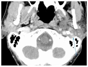
The authors postulate that in this patient the hemifacial spasm commenced with the onset of the malignant transformation in the recurrent pleomorphic adenoma in the parotid gland. In the absence of any evidence of perineural invasion, we believe that peri-tumoral inflammatory responses caused the neural stimulation that resulted in hemifacial spasm. This patient did not have any prior ear surgery or any other known etiology to account for this symptom. An alternative explanation to the patient’s neurological symptoms is external compression to the facial nerve. This could be related to the dense fibrotic tissue surrounding both tumor nodules and nerves or to direct tumor extension into the left stylomastoid foramen[14] (Figure (Figure3).3). The latter mechanism has been previously proposed by Blevins et al[14].
In conclusion, we present the first case of hemifacial spasm in conjunction with transformation of a recurrent pleomorphic adenoma into a carcinoma ex pleomorphic adenoma. The pathophysiology of hemifacial spasms is discussed.
ACKNOWLEDGMENTS
The authors would like to acknowledge the generous support of this research by the Mount Sinai Health System and the THANC Foundation.
COMMENTS
Case characteristics
A 56-year-old female with a history of recurrent pleomorphic adenoma of the left parotid gland treated with surgery and external beam radiation therapy presented with ipsilateral hemifacial spasm.
Clinical diagnosis
The clinical diagnosis is a malignant change in a parotid pleomorphic adenoma with involvement of the facial nerve.
Differential diagnosis
The differential diagnosis is the stimulation of facial nerve by perineural invasion or an inflammatory reaction caused by malignant parotid tumor.
Imaging diagnosis
Repeat CT scan showed enlargement of avidly and uniformly enhancing solid tumor without areas of necrosis or extracapsular extension with extension into the left stylomastoid foramen, along with suspicious changes in enlarged (15 mm) left level IV lymph node (Figure (Figure1A1A).
Pathological diagnosis
Fine-needle aspiration biopsy of the tumor was suspicious for carcinoma ex pleomorphic adenoma. Final surgical pathology confirmed a 5.2 cm pleomorphic adenoma with a multinodular growth pattern with two foci of early non-invasive carcinoma and no malignant spread to adjacent fibroadipose tissue, nerves or thirteen level II-V lymph nodes.
Treatment
A radical parotidectomy with facial nerve sacrifice, ipsilateral selective neck dissection (levels I-IV), and a de-epithelialized anterolateral thigh free flap was performed. A sural nerve grafting from the main trunk of the facial nerve to its branches and an oral commissure suspension with a fascia lata sling was done.
Experiences and lessons
The authors postulate that the hemifacial spasm commenced with the onset of the malignant transformation in the recurrent pleomorphic adenoma in the ipsilateral parotid gland. In the absence of any evidence of perineural invasion, they believe that peri-tumoral inflammatory responses caused the neural stimulation that resulted in hemifacial spasm.
Peer-review
This is the first reported case of malignant transformation of a recurrent pleomorphic adenoma in a parotid gland presenting with ipsilateral hemifacial spasm. In the absence of evidence of perineural invasion of the ipsilateral facial nerve, it is postulated that peri-tumoral inflammatory responses were responsible for the excitation of this nerve and the resultant hemifacial spasm.
Footnotes
Institutional review board statement: This case report was exempt from the Institutional Review Board standards at Mount Sinai Beth Israel in New York.
Informed consent statement: This case report was exempt from obtaining informed consent based on Institutional Review Board standards at Mount Sinai Beth Israel in New York.
Conflict-of-interest statement: All the authors have no conflict of interests to declare.
Manuscript source: Invited manuscript
Specialty type: Oncology
Country of origin: United States
Peer-review report classification
Grade A (Excellent): 0
Grade B (Very good): B
Grade C (Good): C, C
Grade D (Fair): 0
Grade E (Poor): 0
Peer-review started: October 28, 2016
First decision: December 1, 2016
Article in press: January 3, 2017
P- Reviewer: Sedassari BT, Schneider S, Takahashi H S- Editor: Qi Y L- Editor: A E- Editor: Wu HL
References
1. Flint PW. Cummings Otolaryngology- Head and Neck Surgery. Philadelphia: Mosby Elsevier; 2010. [Google Scholar]
2. Antony J, Gopalan V, Smith RA, Lam AK. Carcinoma ex pleomorphic adenoma: a comprehensive review of clinical, pathological and molecular data. Head Neck Pathol. 2012;6:1–9. [PMC free article] [PubMed] [Google Scholar]
3. Destee A, Bouchez B, Pellegrin P, Warot P. Hemifacial spasm associated with a mixed benign parotid tumour. J Neurol Neurosurg Psychiatry. 1985;48:189–190. [PMC free article] [PubMed] [Google Scholar]
4. Zbären P, Tschumi I, Nuyens M, Stauffer E. Recurrent pleomorphic adenoma of the parotid gland. Am J Surg. 2005;189:203–207. [PubMed] [Google Scholar]
5. Seifert G. Histopathology of malignant salivary gland tumours. Eur J Cancer B Oral Oncol. 1992;28B:49–56. [PubMed] [Google Scholar]
6. Witt RL, Eisele DW, Morton RP, Nicolai P, Poorten VV, Zbären P. Etiology and management of recurrent parotid pleomorphic adenoma. Laryngoscope. 2015;125:888–893. [PubMed] [Google Scholar]
7. Boukheris H, Ron E, Dores GM, Stovall M, Smith SA, Curtis RE. Risk of radiation-related salivary gland carcinomas among survivors of Hodgkin lymphoma: a population-based analysis. Cancer. 2008;113:3153–3159. [PMC free article] [PubMed] [Google Scholar]
8. Nouraei SA, Hope KL, Kelly CG, McLean NR, Soames JV. Carcinoma ex benign pleomorphic adenoma of the parotid gland. Plast Reconstr Surg. 2005;116:1206–1213. [PubMed] [Google Scholar]
9. Olsen KD, Lewis JE. Carcinoma ex pleomorphic adenoma: a clinincopathological review. Head Neck. 2001;23:705–712. [PubMed] [Google Scholar]
10. Castiglione M, Broggi M, Cordella R, Acerbi F, Ferroli P. Immediate disappearance of hemifacial spasm after partial removal of ponto-medullary junction anaplastic astrocytoma: case report. Neurosurg Rev. 2015;38:385–390; discussion 390. [PubMed] [Google Scholar]
11. Behbehani R, Hussain AE, Hussain AN. Parotid tumor presenting with hemifacial spasm. Ophthal Plast Reconstr Surg. 2009;25:141–142. [PubMed] [Google Scholar]
12. Yaltho TC, Jankovic J. The many faces of hemifacial spasm: differential diagnosis of unilateral facial spasms. Mov Disord. 2011;26:1582–1592. [PubMed] [Google Scholar]
13. Tan NC, Chan LL, Tan EK. Hemifacial spasm and involuntary facial movements. QJM. 2002;95:493–500. [PubMed] [Google Scholar]
14. Blevins NH, Jackler RK, Kaplan MJ, Boles R. Facial paralysis due to benign parotid tumors. Arch Otolaryngol Head Neck Surg. 1992;118:427–430. [PubMed] [Google Scholar]

