File:Cavernoma Pineale con indicazione.jpg
Size of this preview: 602 × 599 pixels. Other resolution: 1,915 × 1,906 pixels.
Original file (1,915 × 1,906 pixels, file size: 624 KB, MIME type: image/jpeg)
Go to top
Summary
Dettagli
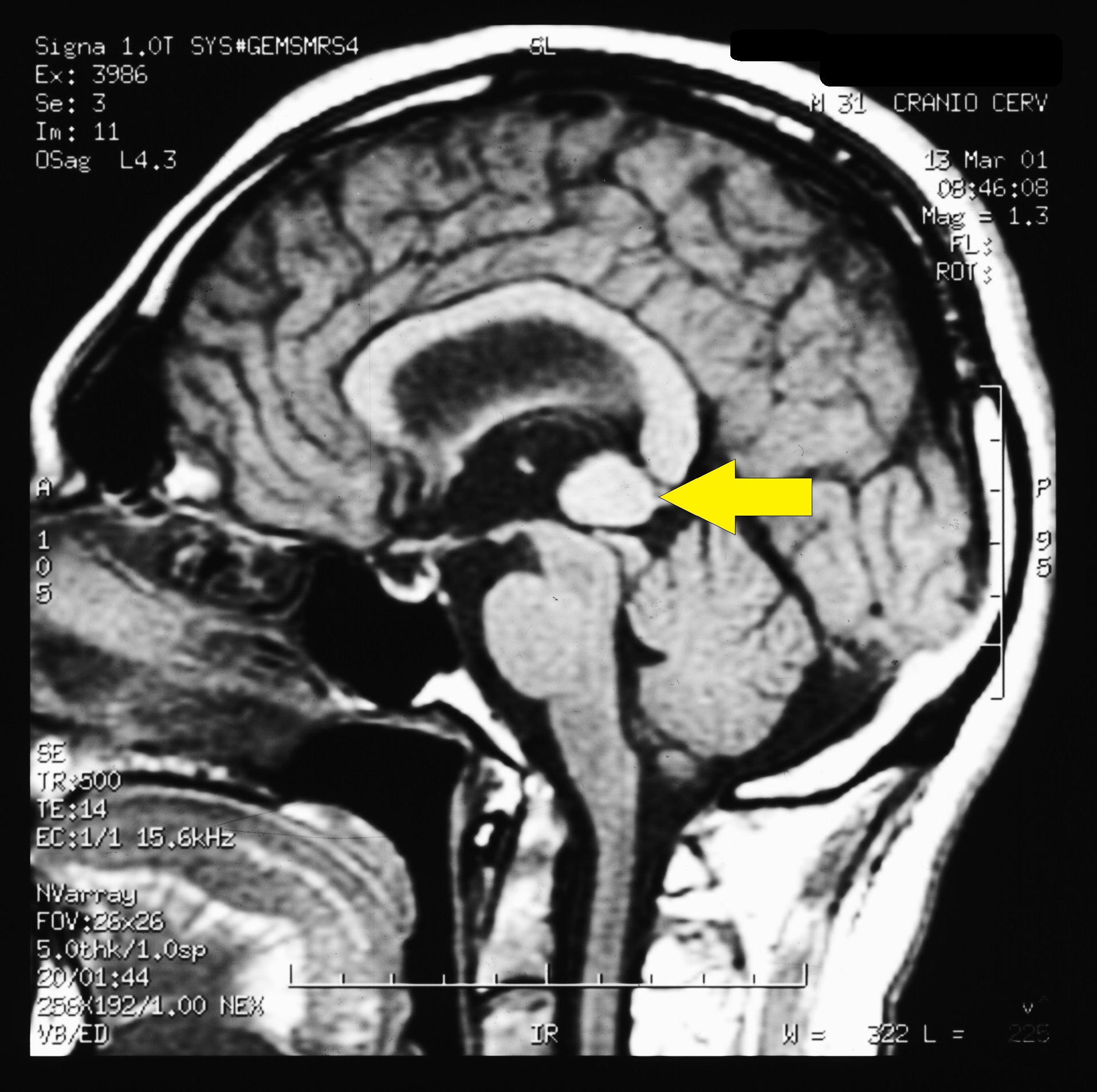
| Description | Brain MR image in bruxist patient affected by Pineal Cavernoma The MR image of the brain performed after intravenous administration of contrast medium in the TSE, FLAIR and GE sequences on the sagittal planes showed the presence of a rounded area of about 1.5 cm in diameter located at the Galeno cistern in a patient with Pineal Cavernoma | ||
|---|---|---|---|
| Source | Archive Studio Frisardi
| ||
| Date | |||
| Author | Gianni Frisardi | ||
| Licensing |
|
Free image from the
You can freely use it as long as you respect the terms of the Creative Commons Attribuzione-Condividi allo stesso modo (CC BY-SA 4.0) license.
Hence, you must always indicate the image's source; in this case it would be enough to link the present page:
https://wiki.masticationpedia.org/index.php/File:Cavernoma_Pineale_con_indicazione.jpg
Archive Studio Frisardi
collection.You can freely use it as long as you respect the terms of the Creative Commons Attribuzione-Condividi allo stesso modo (CC BY-SA 4.0) license.
Hence, you must always indicate the image's source; in this case it would be enough to link the present page:
https://wiki.masticationpedia.org/index.php/File:Cavernoma_Pineale_con_indicazione.jpg
File history
Click on a date/time to view the file as it appeared at that time.
| Date/Time | Thumbnail | Dimensions | User | Comment | |
|---|---|---|---|---|---|
| current | 19:19, 15 February 2022 | | 1,915 × 1,906 (624 KB) | Admin (talk | contribs) | == Dettagli == {{CF | Descrizione = Brain MR image in bruxist patient affected by Pineal Cavernoma <br>''The MR image of the brain performed after intravenous administration of contrast medium in the TSE, FLAIR and GE sequences on the sagittal planes showed the presence of a rounded area of about 1.5 cm in diameter located at the Galeno cistern in a patient with Pineal Cavernoma'' | Fonte = {{SF}} | Data = | Autore = Gianni Frisardi | Licenza = {{Cc-by-sa-4.0}} }} [[Category:Magnetic reson... |
You cannot overwrite this file.
File usage
There are no pages that use this file.