Category:Images provided by Studio Frisardi
Revision as of 17:03, 15 February 2022 by Admin (talk | contribs) (Created page with "Category:Images by source")
Go to top
Pages in category "Images provided by Studio Frisardi"
The following 2 pages are in this category, out of 2 total.
Media in category "Images provided by Studio Frisardi"
The following 68 files are in this category, out of 68 total.
- 5° Caso Clinico- Disfunzione Temporomandibolare.jpg 2,819 × 1,880; 4.82 MB
- Anatomical dissection of the Temporomandibular Joint (TMJ) - Sagittal.jpg 2,048 × 2,739; 3.8 MB
- Aspetto frontale del caso clinico finalizzato.jpg 3,008 × 2,000; 1.21 MB
- Assiografia dx.jpg 800 × 532; 73 KB
- Assiografia sn.jpg 800 × 573; 75 KB
- Atm1 sclerodermia.jpg 379 × 179; 20 KB
- Axio dx.jpg 3,008 × 2,000; 1.48 MB
- Axio sn.jpg 3,008 × 2,000; 1.45 MB
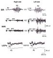
- Bilateral Electric Transcranial Stimulation.jpg 800 × 434; 57 KB
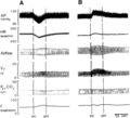
- Bilateral Root-MEPs.jpg 800 × 592; 71 KB
- Caso clinico di 2° Classe con deep bite (3).jpg.jpg 800 × 196; 26 KB
- Caso clinico di 2° Classe con deep bite (4).jpg 799 × 293; 36 KB
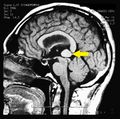
- Cavernoma Pineale con indicazione.jpg 1,915 × 1,906; 624 KB
- Chirurgia Ortognatica.jpg 800 × 531; 42 KB
- Cicli masticatori.jpg 2,559 × 1,526; 299 KB
- CR MIR masseter inhibitory recovery cycle reflex.jpg 800 × 480; 72 KB
- Crossbite.jpg 706 × 471; 38 KB
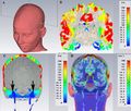
- FEM.jpg 707 × 600; 75 KB
- Final left view of Invisible OrthoNeuroGnathodontic treatments (mirror).jpg 3,008 × 2,000; 1.51 MB
- Final right view of Invisible OrthoNeuroGnathodontic treatments.jpg 3,008 × 2,000; 1.54 MB
- Francesca Frisardi.jpg 455 × 650; 30 KB
- Frontal Occlusal view in TMDs patient with Postpolio Syndrome.png 768 × 512; 939 KB
- Frontal view of Invisible OrthoNeuroGnathodontic treatments.jpg 3,008 × 2,000; 1.55 MB
- Gianni Frisardi mod.jpg 307 × 252; 55 KB
- Gianni Frisardi.jpg 547 × 861; 67 KB
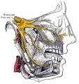
- Gray778 Trigeminal.png 563 × 599; 424 KB
- H-wave.jpg 2,324 × 1,062; 270 KB
- Interference pattern.jpg 1,674 × 860; 380 KB
- Invisible OrthoNeuroGnathodontic before treatments (left side).jpg 2,769 × 1,635; 820 KB
- Invisible OrthoNeuroGnathodontic before treatments (right side).jpg 3,008 × 2,000; 1.06 MB
- Invisible OrthoNeuroGnathodontic before treatments.jpg 3,008 × 2,000; 1.53 MB
- Irene Minciacchi.jpg 465 × 700; 44 KB
- Jaw Jerk .jpg 1,920 × 1,042; 409 KB
- Jaw Jerk in OrtoNeuroGnatodonzia 1.jpg 1,924 × 1,016; 382 KB
- Kreceptor.jpg 1,512 × 1,376; 283 KB
- Laser Evoked Potentials - Blink reflex - Masseter Silent Period.jpg 551 × 599; 61 KB
- Laser test.jpg 700 × 376; 108 KB
- Mechanic Silent Period.jpg 800 × 434; 71 KB
- MGSTIM wiki.jpg 2,925 × 1,950; 3.52 MB
- MIR2.jpg 3,088 × 2,100; 735 KB
- Muscoli masticatori.jpg 959 × 510; 108 KB
- Occlusal Centric view in open and cross bite patient.jpg 706 × 471; 38 KB
- Occlusal view of Invisible OrthoNeuroGnathodontic treatments.jpg 2,420 × 1,906; 701 KB
- Open bite Jaw jerk.jpg 437 × 350; 53 KB
- OrtoNeuroGnatodonzia 1.jpg 3,008 × 2,000; 2.67 MB
- OrtoNeuroGnatodonzia 2.jpg 2,730 × 1,395; 1.77 MB
- OrtoNeuroGnatodonzia 3.jpg 3,008 × 2,000; 2.58 MB
- Periodo Silente in OrtoNeuroGnatodonzia 1.jpg 1,913 × 985; 551 KB
- Potenziale Evocato della Radice Trigeminale.jpg 378 × 220; 28 KB
- Pre Crossbite.jpg 1,940 × 1,279; 599 KB
- PSM - Masseter mechanical Silent Period.jpg 800 × 571; 75 KB
- Pz.Arc001.jpg 768 × 512; 88 KB
- Pz.Arc006.jpg 388 × 599; 28 KB
- Relazione Centrica Neuro Evocata.jpg 3,072 × 2,048; 4.49 MB
- Riflesso mandibolare.jpg 800 × 480; 57 KB
- Silent Period Postpolio.jpg 2,780 × 1,641; 1.67 MB
- Spasmo emimasticatorio assiografia.jpg 2,952 × 1,941; 2.13 MB
- Spasmo emimasticatorio ATM.jpg 477 × 521; 45 KB
- Spasmo emimasticatorio JJ.jpg 699 × 459; 132 KB
- Spasmo emimasticatorio SP.jpg 697 × 427; 150 KB
- Spasmo emimasticatorio TC.jpg 512 × 768; 60 KB
- Spasmo emimasticatorio.jpg 726 × 485; 132 KB
- The phases of paradigm change according to Thomas Kuhn.jpg 203 × 492; 22 KB
- Trigeminal Cortical Area - C-MEPs.jpg 800 × 517; 50 KB
- VEMP.jpg 600 × 298; 17 KB


