File list
This special page shows all uploaded files.
| Date | Name | Thumbnail | Size | User | Description | Versions |
|---|---|---|---|---|---|---|
| 08:32, 7 February 2022 | Logo2.png (file) |  |
16 KB | Admin | 1 | |
| 16:47, 15 February 2022 | Wiki.png (file) |  |
5 KB | Admin | == Summary == Importing files from prev WIKI Category:Masticationpedia logo | 1 |
| 16:48, 15 February 2022 | 5.png (file) |  |
793 KB | Admin | <noinclude>Category:Unsorted files</noinclude> | 1 |
| 16:53, 15 February 2022 | 8C06B0F5-086E-4F1C-9FF0-6A988232C556.jpg (file) |  |
114 KB | Admin | <noinclude>Category:Unsorted files</noinclude> | 1 |
| 16:53, 15 February 2022 | 5° Caso Clinico- Disfunzione Temporomandibolare.jpg (file) |  |
4.82 MB | Admin | ==Details== {{CF | Descrizione = Electrode provision in the neuro Evoked Occlusal Centric Relation on TMDs patient<br> ''Electrode provision in the neuro Evoked Occlusal Centric Relation on TMDs patient. Cathodic and recording electrodes located on parietal bone and masseter muscles, respectively'' | Fonte = {{SF}} | Data = | Autore = {{Augf}} | Licenza = {{Cc-by-sa-4.0}} }} Category:Patients | 1 |
| 16:58, 15 February 2022 | A7A6C3FB-BB01-4F06-9CA2-0EC83D6A1A68.jpeg (file) | 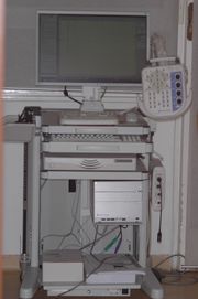 |
402 KB | Admin | Category:Generic files | 1 |
| 17:01, 15 February 2022 | Alice Bisirri.jpg (file) |  |
29 KB | Admin | 1 | |
| 17:03, 15 February 2022 | Anatomical dissection of the Temporomandibular Joint (TMJ) - Sagittal.jpg (file) |  |
3.8 MB | Admin | == Dettagli == {{CF | Descrizione = Sagittal aspect of an anatomical dissection of the Temporomandibular Joint (TMJ) | Fonte = {{SF}} | Data = | Autore = {{Augf}} | Licenza = {{Cc-by-sa-4.0}} }} | 1 |
| 17:05, 15 February 2022 | Andrea Melcarne.jpg (file) | 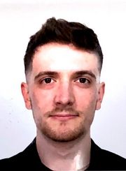 |
50 KB | Admin | <noinclude>Category:Unsorted files</noinclude> | 1 |
| 17:05, 15 February 2022 | Andrea Melcarne CV.pdf (file) | 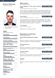 |
135 KB | Admin | <noinclude>Category:Unsorted files</noinclude> | 1 |
| 17:06, 15 February 2022 | Artur-tumasjan-qLzWvcQq-V8-unsplash.jpg (file) |  |
46 KB | Admin | {{US | Descrizione = an apple a day keeps the doctor away<!-- inserisci qui tutti i termini che identificano il concetto che l'immagine spiega --> | Fonte = derived from [https://unsplash.com/photos/qLzWvcQq-V8 this picture] - [https://www.unsplash.com unsplash.com] <!-- da dove viene il file? --> | Data = <!-- se nota, data di scatto dell'immagine o di creazione del file --> | Autore = [https://unsplash.com/@arturtumasjan?utm_source=unsplash&utm_medium=referral&utm_content=creditCopyText Art... | 1 |
| 17:09, 15 February 2022 | Aspetto frontale del caso clinico finalizzato.jpg (file) |  |
1.21 MB | Admin | == Dettagli == {{CF | Descrizione = Frontal aspect post OrthoNeuroGnathodontics treatment<br>''Frontal aspect post treatment in a 2° Class facial hypodivergent morphology (5 years of follow up), treated through OrthoNeuroGnathodontics '' | Fonte = {{SF}} | Data = | Autore = {{auff}} | Licenza = {{Cc-by-sa-4.0}} }} Category:Clinical cases Category:OrthoNeuroGnatodontics | 1 |
| 17:55, 15 February 2022 | Assiografia dx.jpg (file) | 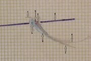 |
73 KB | Admin | == Dettagli == {{CF | Descrizione = ''Right Axiographic traces performed with paraocclusal clutches (003) in a healthy subject. <br> a) Condylar rotation center; <br> b) Phonetic area; <br> c) Laterotrusive condylar tracking; <br> d) Orbital-axis plane; <br> e) Protrusive condylar tracking; <br> f) Mediotrusive condylar tracking;<br> g) Mediotrusive functional area of the masticatory cycles; <br> h) Laterotrusive functional area of the masticatory cycles | Fonte = {{SF}} | Data = | Autore... | 1 |
| 17:55, 15 February 2022 | Assiografia sn.jpg (file) | 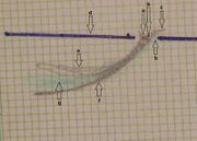 |
75 KB | Admin | == Dettagli == {{CF | Descrizione = Left Axiographic traces performed with paraocclusal clutches (003) in a healthy subject. <br> a) Condylar rotation center; <br> b) Phonetic area; <br> c) Laterotrusive condylar tracking; <br> d) Orbital-axis plane; <br> e) Protrusive condylar tracking; <br> f) Mediotrusive condylar tracking; <br> g) Mediotrusive functional area of the masticatory cycles; <br> h) Laterotrusive functional area of the masticatory cycles | Fonte = {{SF}} | Data = | Autore =... | 1 |
| 17:56, 15 February 2022 | Atm1 sclerodermia.jpg (file) |  |
20 KB | Admin | {{SF}} Category:Tomography Category:Scleroderma | 1 |
| 18:00, 15 February 2022 | Axio dx.jpg (file) | 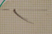 |
1.48 MB | Admin | == Details == {{CF | Descrizione = ''Right Axiographic traces performed with paraocclusal clutches (002) in a patient with Pineal Cavernoma'' | Fonte = {{SF}} | Data = | Autore = Gianni Frisardi | Licenza = {{Cc-by-sa-4.0}} }} Category:Axiography | 1 |
| 18:01, 15 February 2022 | Axio sn.jpg (file) | 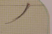 |
1.45 MB | Admin | == Dettagli == {{CF | Descrizione = Left Axiographic traces performed with paraocclusal clutches (001)<br>''Left Axiographic traces performed with paraocclusal clutches in a patient with Pineal Cavernoma'' | Fonte = {{SF}} | Data = | Autore = Gianni Frisardi | Licenza = {{Cc-by-sa-4.0}} }} Category:Axiography | 1 |
| 18:01, 15 February 2022 | B2 Stealth formation.jpg (file) | 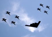 |
653 KB | Admin | {{fileinfo | Descrizione = Valiant Shield - B2 Stealth bomber from Missouri leads ariel formation<!-- inserisci qui tutti i termini che identificano il concetto che l'immagine o il file spiegano --> | Fonte = derived from [https://commons.wikimedia.org/wiki/File:Valiant_Shield_-_B2_Stealth_bomber_from_Missouri_leads_ariel_formation.jpg B2 Stealth bomber from Missouri, Public domain, via Wikimedia Commons] <!-- da dove viene il file? --> | Data = 2006-2021<!-- data di scatto dell'immagine o di... | 1 |
| 18:02, 15 February 2022 | Bilateral Electric Transcranial Stimulation.jpg (file) |  |
57 KB | Admin | == Details == {{CF | Descrizione = Motor Evoked Potential in hemilateral occlusal crossbite patient<br>''Motor Evoked Potential by electrical Transcranial Stimulation of the trigeminal roots. Note the structural symmetry calculated by the peak-to-peak amplitude on the right (upper trace) and left masseters (lower trace). (Nemus2, NGF; EBNeuro, Firenze, Italy)'' | Fonte = {{SF}} | Data = | Autore = Gianni Frisardi | Licenza = {{Cc-by-sa-4.0}} }} | 1 |
| 18:02, 15 February 2022 | Bilateral Magnetic Transcranial Stimulation in the masticatory rehabilitation.jpg (file) |  |
1.41 MB | Admin | == Dettagli == {{CF | Descrizione = ''Bilateral Magnetic Transcranial Stimulation in the masticatory rehabilitation procedures ([https://www.esaote.com/it-IT/ Esaote, Genoa, Italy])'' | Fonte = {{SF}} | Data = | Autore = {{Augf}} | Licenza = {{Cc-by-sa-4.0}} }} Category:Patients | 1 |
| 18:03, 15 February 2022 | Bilateral Root-MEPs.jpg (file) | 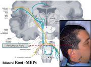 |
71 KB | Admin | ==Dettagli== {{CF | Descrizione = ''electrical Transcranial Stimulation: Schematic and anatomical representation.'' | Fonte = {{SF}} | Data = | Autore = {{augf}} | Licenza = {{Cc-by-sa-4.0}} }} Category:Explanatory files | 1 |
| 18:03, 15 February 2022 | Boostrapping.jpg (file) |  |
65 KB | Admin | <noinclude>Category:Unsorted files</noinclude> | 1 |
| 18:04, 15 February 2022 | Brainstorming session.jpg (file) |  |
51 KB | Admin | == Details == {{US | Descrizione = Brainstorming session<!-- inserisci qui tutti i termini che identificano il concetto che l'immagine spiega --> | Fonte = [https://www.unsplash.com unsplash.com] https://unsplash.com/photos/IBUcu_9vXJc <!-- da dove viene il file? --> | Data = <!-- se nota, data di scatto dell'immagine o di creazione del file --> | Autore = [https://unsplash.com/@thomasdrouaultphotography?utm_source=unsplash&utm_medium=referral&utm_content=creditCopyText Thomas Drouault] | Lic... | 1 |
| 18:05, 15 February 2022 | Button.gif (file) |  |
2 KB | Admin | <noinclude>Category:Unsorted files</noinclude> | 1 |
| 18:06, 15 February 2022 | CR MIR masseter inhibitory recovery cycle reflex.jpg (file) |  |
72 KB | Admin | ==Dettagli== {{CF | Descrizione = Representation of the masseter inhibitory recovery cycle reflex<br> ''Note the pair of electrical stimuli (S1 and S2) and the corresponding silent periods (ES1 and ES2). ([https://www.nihonkohden.com/index.html Nihon Koden, Tokio, Japan])'' | Fonte = {{SF}} | Data = | Autore = {{augf}} | Licenza = {{Cc-by-sa-4.0}} }} | 1 |
| 18:07, 15 February 2022 | CV Bisirri Alice EN.pdf (file) | 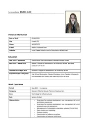 |
234 KB | Admin | <noinclude>Category:Unsorted files</noinclude> | 1 |
| 18:09, 15 February 2022 | Carlo Leonardis.jpg (file) | 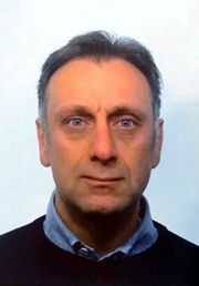 |
24 KB | Admin | Category:Images of Masticationpedia authors | 1 |
| 18:15, 15 February 2022 | London shelton.jpg (file) |  |
81 KB | Admin | Category:Masticationpedia (Charity) | 1 |
| 18:16, 15 February 2022 | Caso clinico di 2° Classe con deep bite (2) - 1st phase of OrthoNeuroGnatodontic treatment (001).jpg (file) |  |
54 KB | Admin | ==Dettagli== {{CF | Descrizione = Lateral view of the 1st phase of OrthoNeuroGnatodontic treatment (001)<br>Class 2 elastics used for avoiding the against mesializing vector on the upper dental frontal group''<br>''Frontal aspect before (left) and post (right) in a 2° Class facial hypodivergent morphology, treated through OrtoNeuroGnathodontics'' | Fonte = {{SF}} | Data = | Autore = Flavio Frisardi | Licenza = {{Cc-by-sa-4.0}} }} Category:OrthoNeuroGnatodontics | 1 |
| 18:17, 15 February 2022 | Caso clinico di 2° Classe con deep bite (3).jpg.jpg (file) | 26 KB | Admin | ==Dettagli== {{CF | Descrizione = Second phase in tne OrthoNeuroGnathodontic 2° Class facial hypodivergent morphology treatment (001) <br>''Occlusal and frontal view of Second phase in tne OrthoNeuroGnathodontic 2° Class facial hypodivergent morphology treatment'' | Fonte = {{SF}} | Data = | Autore = Flavio Frisardi | Licenza = {{Cc-by-sa-4.0}} }} Category:OrthoNeuroGnatodontics | 1 | |
| 18:17, 15 February 2022 | Caso clinico di 2° Classe con deep bite (4).jpg (file) |  |
36 KB | Admin | == Dettagli == {{CF<!--Collezione Frisardi--> | Descrizione = Vertical Occlusal Dimension increase in OrthoNeuroGnathodontic treatments (001)<br>''Increase of Vertical Occlusal Dimension (left side) and Jaw jerk recorded on right masseter (upper trace) and left masseter (lower trace) after 5 years of follow up in a 2° Class facial hypodivergent morphology, treated through OrthoNeuroGnathodontics'' | Fonte = {{SF}} <!-- da dove viene il file? --> | Data = <!-- se nota, data di scatto dell'imma... | 1 |
| 18:18, 15 February 2022 | Caso clinico di 2° Classe con deep bite (5) - facial hypodivergent morphology, OrtoNeuroGnathodontics.jpg (file) |  |
27 KB | Admin | ==Dettagli== {{CF | Descrizione = Frontal view before (left) and post (right) in OrthoNeuroGnathodontic treatment (001)<br>''Frontal aspect before (left) and post (right) in a 2° Class facial hypodivergent morphology and deep bite treated through OrthoNeuroGnathodontics'' | Fonte = {{SF}} | Data = | Autore = {{auff}} | Licenza = {{Cc-by-sa-4.0}} }} Category:Clinical cases Category:OrthoNeuroGnatodontics | 1 |
| 18:19, 15 February 2022 | Cavernoma Pineale con indicazione.jpg (file) | 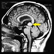 |
624 KB | Admin | == Dettagli == {{CF | Descrizione = Brain MR image in bruxist patient affected by Pineal Cavernoma <br>''The MR image of the brain performed after intravenous administration of contrast medium in the TSE, FLAIR and GE sequences on the sagittal planes showed the presence of a rounded area of about 1.5 cm in diameter located at the Galeno cistern in a patient with Pineal Cavernoma'' | Fonte = {{SF}} | Data = | Autore = Gianni Frisardi | Licenza = {{Cc-by-sa-4.0}} }} [[Category:Magnetic reson... | 1 |
| 18:19, 15 February 2022 | Cesare Iani.jpg (file) |  |
53 KB | Admin | Category:Images of Masticationpedia authors | 1 |
| 18:20, 15 February 2022 | Chirurgia Ortognatica.jpg (file) | 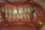 |
42 KB | Admin | == Details == {{CF | Descrizione = ''Occlusal Centric view before neuro Evoked Centric Relation in patient undergoing orthognathic surgery. <br>Note the misalignment of the incisal line'' | Fonte = {{SF}} | Data = | Autore = {{augf}} | Licenza = {{Cc-by-sa-4.0}} }} | 1 |
| 18:20, 15 February 2022 | Cicli masticatori.jpg (file) | 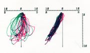 |
299 KB | Admin | ==Dettagli== {{CF | Descrizione = Masticatory Electrognathographic traces.<br>''Coronal and sagittal Electrognathographic view of masticatory cycles, left and right side respectively'' | Fonte = {{SF}} | Data = | Autore = Gianni Frisardi | Licenza = {{Cc-by-sa-4.0}} }} Category:Electromyography Category:Electrognathography | 1 |
| 18:21, 15 February 2022 | Collaborazione Scientifica.jpg (file) |  |
42 KB | Admin | Category:Masticationpedia documents | 1 |
| 18:22, 15 February 2022 | Collaborazione Scientifica.pdf (file) |  |
298 KB | Admin | Category:PDF files Category:Masticationpedia documents | 1 |
| 18:23, 15 February 2022 | Contact your dentist.jpg (file) |  |
808 KB | Admin | {{US | Descrizione = Contact your dentist<!-- inserisci qui tutti i termini che identificano il concetto che l'immagine spiega --> | Fonte = [https://www.unsplash.com unsplash.com] https://unsplash.com/photos/vu-DaZVeny0<!-- da dove viene il file? --> | Data = <!-- se nota, data di scatto dell'immagine o di creazione del file --> | Autore = [https://unsplash.com/@anikolleshi?utm_source=unsplash&utm_medium=referral&utm_content=creditCopyText Ani Kolleshi]<!-- autore dell'immagine/file --> | Li... | 1 |
| 18:24, 15 February 2022 | Crossbite.jpg (file) | 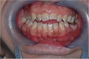 |
38 KB | Admin | ==Details== {{CF | Descrizione = Occlusal Centric view in open bite patient <br> ''Occlusal Centric view in open and cross bite patient '' | Fonte = {{SF}} | Data = | Autore = {{augf}} | Licenza = {{Cc-by-sa-4.0}} }} | 1 |
| 18:25, 15 February 2022 | Dentistry workplace.jpg (file) | 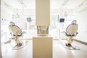 |
942 KB | Admin | {{US | Descrizione = Dentistry workplace<!-- inserisci qui tutti i termini che identificano il concetto che l'immagine spiega --> | Fonte = [https://www.unsplash.com unsplash.com] https://unsplash.com/photos/D0ov97Td-xM<!-- da dove viene il file? --> | Data = <!-- se nota, data di scatto dell'immagine o di creazione del file --> | Autore = [https://unsplash.com/@michaelwb?utm_source=unsplash&utm_medium=referral&utm_content=creditCopyText Michael Browning]<!-- autore dell'immagine/file --> | L... | 1 |
| 18:27, 15 February 2022 | Diego Centonze.jpg (file) |  |
18 KB | Admin | Category:Images of Masticationpedia authors | 1 |
| 18:28, 15 February 2022 | Dummy.pdf (file) |  |
13 KB | Admin | Category:Dummy contents | 1 |
| 18:29, 15 February 2022 | EBNeuro.jpg (file) | 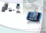 |
317 KB | Admin | Category:Generic files | 1 |
| 18:30, 15 February 2022 | Electrical stimulation of cranial nerves in cognition and disease.pdf (file) |  |
3.4 MB | Admin | {{Fresinfo | Descrizione = [https://www.brainstimjrnl.com/action/showPdf?pii=S1935-861X%2820%2930041-3 Electrical stimulation of cranial nerves in cognition and disease] - [http://crossmark.crossref.org/dialog/?doi=10.1016/j.brs.2020.02.019&domain=pdf updates]<!-- inserisci qui tutti i termini che identificano il concetto che l'immagine o il file spiegano --> | Fonte = https://doi.org/10.1016/j.brs.2020.02.019<!-- da dove viene il file? --> | Data = 18 July 2019<!-- data di scatto dell'immagi... | 1 |
| 18:31, 15 February 2022 | Evans2.png (file) |  |
27 KB | Admin | == Summary == {{Informazioni file | Descrizione = Image from Resources:Measuring Statistical Evidence Using Relative Belief | Fonte = https://pubmed.ncbi.nlm.nih.gov/26925207/ | Data = Retrieved 17/06/2020<!-- dd/mm/yyyy --> | Autore = [[Author:Michael Evans{{!}}Michael Evans]] | Licenza = CC BY | Altre versioni = }} Category:Mathematical expressions | 1 |
| 18:31, 15 February 2022 | Evans3.png (file) | 12 KB | Admin | == Summary == {{Informazioni file | Descrizione = Image from Resources:Measuring Statistical Evidence Using Relative Belief | Fonte = https://pubmed.ncbi.nlm.nih.gov/26925207/ | Data = Retrieved 17/06/2020<!-- dd/mm/yyyy --> | Autore = [[Author:Michael Evans{{!}}Michael Evans]] | Licenza = CC BY | Altre versioni = }} Category:Mathematical expressions | 1 | |
| 18:32, 15 February 2022 | Evans4.png (file) | 14 KB | Admin | == Summary == {{Informazioni file | Descrizione = Image from Resources:Measuring Statistical Evidence Using Relative Belief | Fonte = https://pubmed.ncbi.nlm.nih.gov/26925207/ | Data = Retrieved 17/06/2020<!-- dd/mm/yyyy --> | Autore = [[Author:Michael Evans{{!}}Michael Evans]] | Licenza = CC BY | Altre versioni = }} Category:Mathematical expressions | 1 | |
| 18:33, 15 February 2022 | Evans5.png (file) | 12 KB | Admin | == Summary == {{Informazioni file | Descrizione = Image from Resources:Measuring Statistical Evidence Using Relative Belief | Fonte = https://pubmed.ncbi.nlm.nih.gov/26925207/ | Data = Retrieved 17/06/2020<!-- dd/mm/yyyy --> | Autore = [[Author:Michael Evans{{!}}Michael Evans]] | Licenza = CC BY | Altre versioni = }} Category:Mathematical expressions | 1 | |
| 18:34, 15 February 2022 | Evans6.png (file) | 13 KB | Admin | == Summary == {{Informazioni file | Descrizione = Image from Resources:Measuring Statistical Evidence Using Relative Belief | Fonte = https://pubmed.ncbi.nlm.nih.gov/26925207/ | Data = Retrieved 17/06/2020<!-- dd/mm/yyyy --> | Autore = [[Author:Michael Evans{{!}}Michael Evans]] | Licenza = CC BY | Altre versioni = }} Category:Mathematical expressions | 1 |