File:FEM.jpg
No higher resolution available.
FEM.jpg (707 × 600 pixels, file size: 75 KB, MIME type: image/jpeg)
Go to top
Summary
Dettagli
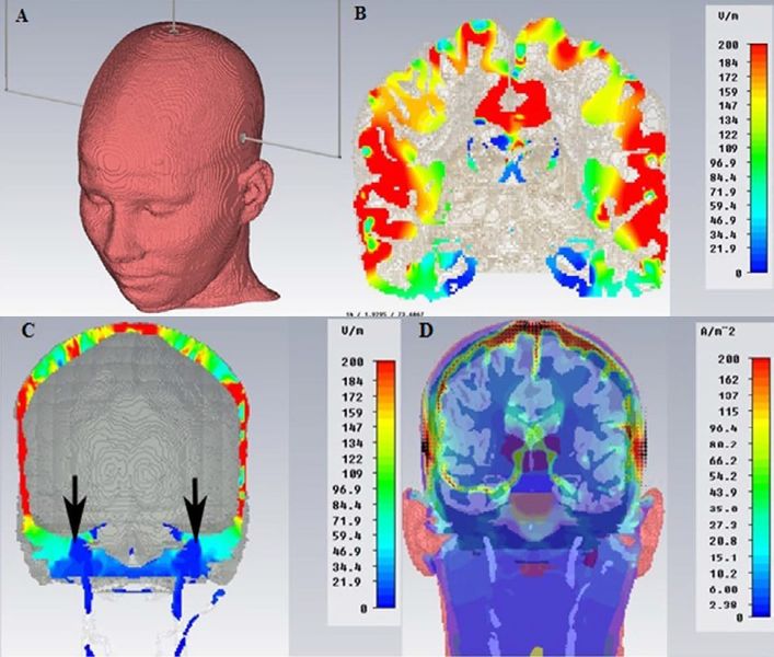
| Description | Finite Elements view of the electric field within the intracranial brain tissue To visualize the distribution of the electric field within the intracranial brain tissue, we report an analysis carried out through a generic finite element processor (FE, SimNibs method). | ||
|---|---|---|---|
| Source | Archive Studio Frisardi
| ||
| Date | |||
| Author | Gianni Frisardi (Studio Frisardi) | ||
| Licensing |
|
Free image from the
You can freely use it as long as you respect the terms of the Creative Commons Attribuzione-Condividi allo stesso modo (CC BY-SA 4.0) license.
Hence, you must always indicate the image's source; in this case it would be enough to link the present page:
https://wiki.masticationpedia.org/index.php/File:FEM.jpg
Archive Studio Frisardi
collection.You can freely use it as long as you respect the terms of the Creative Commons Attribuzione-Condividi allo stesso modo (CC BY-SA 4.0) license.
Hence, you must always indicate the image's source; in this case it would be enough to link the present page:
https://wiki.masticationpedia.org/index.php/File:FEM.jpg
File history
Click on a date/time to view the file as it appeared at that time.
| Date/Time | Thumbnail | Dimensions | User | Comment | |
|---|---|---|---|---|---|
| current | 19:35, 15 February 2022 | | 707 × 600 (75 KB) | Admin (talk | contribs) | ==Dettagli== {{CF | Descrizione = Finite Elements view of the electric field within the intracranial brain tissue<br> ''To visualize the distribution of the electric field within the intracranial brain tissue, we report an analysis carried out through a generic finite element processor (FE, SimNibs method).'' | Fonte = {{SF}} | Data = | Autore = {{augf}} | Licenza = {{Cc-by-sa-4.0}} }} |
You cannot overwrite this file.
File usage
The following 5 pages use this file: